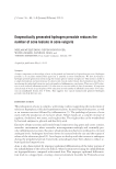
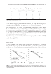
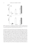
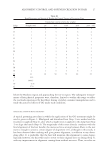
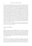
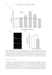
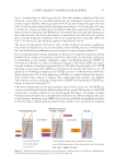
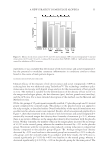
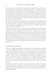
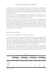
JOURNAL OF COSMETIC SCIENCE 40 Figure 4. Effect of EGCG on antioxidant enzymes SOD and GSH-px and the production of LPO MDA. Data for this statistical analysis were obtained from four independent experiments. *p 0.05 **p 0.01 by Student’s t-test, compared with H2O2 alone.

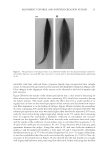
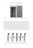
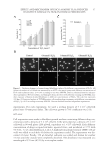
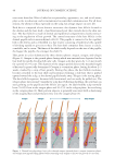
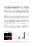
EFFECT AND MECHANISM OF EGCG AGAINST H2O2-INDUCED OXIDATIVE DAMAGE IN HUMAN DERMAL FIBROBLASTS 41 (Nanjing, China). Fibroblasts were plated onto 30-mm petri dishes with a density of 1 × 10⁴ cells/dish, and left overnight. After the cells were completely adherent, they were incubated with H2O2 and H2O2 and EGCG for 6 h, respectively, and the cell protein was extracted. Finally, the contents of SOD, LPO product MDA and GSH-px were determined. DATA ANALYSIS SAS 6.0 was used for statistics analysis. Results were expressed as mean ± standard devia- tion. The analysis of variance (ANOVA) test was used for comparison among groups. The mean values between groups were compared using Student’s t-test. A p value 0.05 indicated a statistical signifi cance. RESULTS AND DISCUSSION H2O2-INDUCED HUMAN DERMAL FIBROBLASTS INJURY AND APOPTOSIS We fi rst used MTT assay to examine the survival rate of H2O2-induced human dermal fi broblasts and established the cell oxidative injury model. The H2O2 treatment time was 3, 6, 12, and 24 h with the concentration of 0.2, 0.4, 0.6, 0.8, 1.0, and 1.2mmol/l. The results showed that the death rate was highly correlated with the concentration of the treatment, and was less correlated with the treatment time. At the same concentration, there is no signifi cant difference among the groups with different treatment time (Figure 1A). Based on the MTT assay results, we chose 6 h and 0.4–0.8mmol/l as the optimal induction conditions for the next series of experiments. Because the MTT assay could not show the kind of cell damage H2O2 treatment caused, we used Hoechst staining to deter- mine whether H2O2-induced fi broblast damage was associated with apoptosis. Hoechst 33342 is a specifi c fl uorescent dye that binds to the minor groove of DNA bases and can detect changes in the nuclear morphology of the apoptotic cells. Under the fl uorescence microscope, the control group showed uniformly stained larger nuclei and evenly dis- persed blue fl uorescence, whereas the H2O2-treated cells showed dense nuclei with brighter fl uorescence in blue and white than normal cells. In the nuclei of the apoptotic cells, dense granular fl uorescence was also observed. They exhibited a typical nuclear condensation and fragmentation, especially under H2O2 concentration of 0.8 mmol/l (Figure 1B, upper panel). We also used the TUNEL method to check the cell apoptosis under a fl uorescence microscope and semiquantitatively determined the rate of cell apop- tosis. The control group showed almost no green fl uorescence, whereas the H2O2-treated group showed positive green fl uorescent cells. The nuclei of the H2O2-treated group showed a typical form of apoptosis with nuclear condensation and fragmentation, and the number of apoptotic cells increased with the H2O2 concentration (Figure 1B, lower panel). In 100× high magnifi cation lens, the number of apoptotic cells randomly selected from fi ve discrete horizons in each group also increased with the H2O2 concentration (Figure 1C). It further confi rmed that H2O2-induced oxidative damage led to apoptosis. H2O2 is a reactive oxygen species with a high reactivity that can pro mote the generation of free radicals, causing membrane LPO, and can freely diffuse in the membrane, result- ing in cell necrosis and apoptosis. The result of the current study further consolidated this conclusion.
Purchased for the exclusive use of nofirst nolast (unknown)
From: SCC Media Library & Resource Center (library.scconline.org)