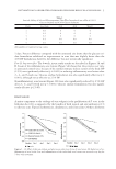
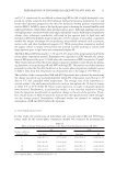
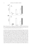
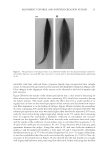
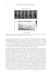
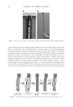
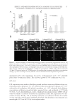
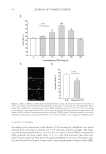
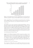
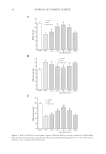
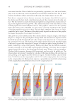
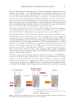
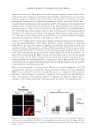
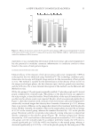
JOURNAL OF COSMETIC SCIENCE 42 EGCG PROTECTS CELLS AGAINST H2O2-INDUCED CELL INJURY AND APOPTOSIS We next studied whether the EGCG-treated human dermal fi broblasts had the ability to fi ght against the oxidative stress by using the H2O2-induced human dermal fi broblasts apoptotic model. We fi rst added EGCG with concentrations ranged from 10 to 200 μg/ml and tested the survival rate of the cells after H2O2 treatment with MTT assay. The results suggested that EGC effectively improved the survival rate of H2O2-treated cells. In particular, they showed signifi cant differences in 20, 50, and 100 μg/ml groups (p 0.05), and the 50 μg/ml group showed the most prominent effect. The statistics from three repeated experiments are shown in Figure 2A. The results from TUNEL assay experiments showed that by adding EGCG (50 μg/ml) the number of positive green fl uo- rescence cells is signifi cantly less than that of the cells treated with H2O2 only (Figure 2B). In 100× high magnifi cation lens, the number of apoptotic cells in the group treated with both H2O2 and EGCG was signifi cantly less than that in the group treated with H2O2 alone (p 0.05, Figure 1C). The above results indicated that EGCG had a protective effect against the H2O2-induced oxidative stress injury in human dermal fi broblasts. EFFECT OF EGCG ON DPPH RADICAL SCAVENGING We further studied whether EGCG has the ability to scavenge the free radicals using the DPPH radical spectrophotometric analysis. The effects of different concentrations of EGCG on free radical scavenging are shown in Figure 3. The results showed that with a concentration range of 1–200 μg/ml, EGCG had the ability to scavenge the free radicals and its effect increased as concentrations increased. With 200 μg/ml EGCG, its effect on radical scavenging reached up to 91.42%. Thus, EGCG has signifi cant effects on free radical scavenging. EFFECT OF EGCG ON SOD, GSH-PX ACTIVITY, AND MDA LEVEL OF HUMAN DERMAL FIBROBLASTS Lipid peroxide-mediated cell damage is one of the major causes of cell injury. MDA is the main product of LPO, and its level indirectly refl ects the degree of oxidative damage to cells. MDA level is also widely used as a biomarker for oxidative stress (16). In addition, antioxidant enzymes such as SOD and GSH-px are also considered to be effective in en- hancing the cellular antioxidant defense system (17). Therefore, we further explored the effects of EGCG on its protection against H2O2-induced cell oxidative stress and apopto- sis by measuring SOD, GSH-px activity, and MDA levels. The results showed that H2O2 dramatically reduced the SOD level in the human dermal fi broblasts compared to the control group. After adding EGCG, the SOD level could be signifi cantly recovered. Moreover, within the concentration range of 10–50 μg/ml, the effect of EGCG on recov- ery of SOD level was found to be concentration-dependent (Figure 4A). When the EGCG concentrations exceeded 50 μg/ml, the ability of EGCG on the recovery of SOD reduced and showed no signifi cant difference compared to the group treated with H2O2 alone (Figure 4A).This may result from the high concentration of EGCG-induced cell toxicity. EGCG also has effect on increasing the activity of GSH-px, which is another antioxidant enzyme. As shown in Figure 4B, H2O2 decreased the GSH-px activity of human dermal fi broblasts, and after adding EGCG, the GSH-px activity can be signifi cantly recovered. Similarly, the recovery of GSH-px activity showed a concentration-dependent effect when the EGCG concentration was in the range of 10–50 μg/ml. When the EGCG concentration

EFFECT AND MECHANISM OF EGCG AGAINST H2O2-INDUCED OXIDATIVE DAMAGE IN HUMAN DERMAL FIBROBLASTS 43 exceeded 50 μg/ml, the ability of EGCG on the recovery of GSH-px activity also de- creased, showing no signifi cant difference compared to the group treated with H2O2 alone (Figure 4B). This result further supported the cell toxicity caused by the high con- centration of EGCG. Similar conclusions were also obtained from the experiments of MDA level measure- ments. As shown in Figure 4C, H2O2 increased the MDA level of human dermal fi bro- blasts and after adding EGCG, the increased MDA level was signifi cantly reduced. However, this effect disappeared under high concentrations of EGCG. In summary, our results showed that EGCG can increase endogenous antioxidant enzymes activity (SOD and GSH-px) and can enhance the OFR scavenging capacity of human body’s own antioxidant defense system while reducing the MDA level. CONCLUSIONS In this study, for the fi rst time we have established the H2O2-induced human dermal fi broblasts as an oxidative injury model. By using different methods such as MTT assay, Hoechst staining, and the in situ TUNEL assay, we confi rmed that H2O2 could inhibit the viability of human dermal fi broblasts and induced the apoptosis at a certain concen- tration. EGCG showed a good protective antioxidative effect and signifi cantly inhibited the oxidation-induced apoptosis in the TUNEL experiments. We also tried to detect the apoptosis by using fl ow cytometer. However, the result is not as convincing as the TUNEL experiments. This study also confi rmed that EGCG increases SOD and GSH-px activity in human dermal fi broblasts and lowers the MDA level, signifi cantly inhibits the LPO in the human dermal fi broblasts, and shows a clear protective effect against H2O2 damage. Therefore, this method provides a theoretical basis for the application of EGCG in cos- metics and is a simple, fast way to screen the cosmetics with antioxidant effect. This is also worthy of further research and be a useful application in terms of screening cosmetics raw materials. ACKNOWLEDGMENT The authors thank Dr. Yan-Ai Mei, professor at School of Life Sciences, Fudan University, for valuable comments on the manuscript. REFERENCES (1) J. H. Chung, S. H. Youn, O. S. Kwon, H. C. Eun, K. H. Kim, K. C. Park, K. H. Cho, and J. I. Youn, Enhanced proliferation and collagen synthesis of human dermal fi broblasts in chronically photodam- aged skin, Photodermatol. Photoimmunol. Photomed., 12, 84–89 (1996). (2) H. G. Vogel, Age-dependent changes in skin biomechanics, measurements in vitro and in vivo. Z. Gerontol., 27, 182–185 (1994). (3) Y. Nishimori, C. Edwards, A. Pearse, K. Matsumoto, M. Kawai, and R. Marks, Degenerative alterations of dermal collagen fi ber bundles in photodamaged human skin and UV-irradiated hairless mouse skin: Possible effect on decreasing skin mechanical properties and appearance of wrinkles, J. Invest. Dermatol., 117, 1458–1463 (2001).
Purchased for the exclusive use of nofirst nolast (unknown)
From: SCC Media Library & Resource Center (library.scconline.org)