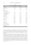
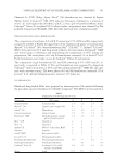
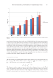
JOURNAL OF COSMETIC SCIENCE 344 85°C and SG (1 g) was added. The melted lipid phase was dispersed in the hot (85°C) surfactant solution (Pluronic® F68, 1.5 g) by using a high-speed stirrer (UltraTurrax T25 IKA-Werke GmbH & Co. KG, Staufen, Germany) at 8000 rpm. The obtained preemul- sion was ultrasonifi ed by using a UP 400 S (Ultraschallprozessor, Dr. Hielscher GmbH, Teltow, Germany) maintaining the temperature at least 5°C above the lipid melting point. After US method, the obtained dispersion was cooled in an ice bath to solidify the lipid matrix and to form SLNs. To acquire insights about the mechanism involving SG release from SLN, we prepared two different hydrogels (SLN-IN and SLN-OUT) using glycerol and xanthan gum as excipients (12). Briefl y, SLN-IN formulation was produced adding to SG-loaded SLN suspensions (89%, w/w), 10% (w/w) of glycerol, and 1% (w/w) of xanthan gum, whereas SLN-OUT was produced adding to a suspension of not loaded SLN and free SG (89%, w/w), 10 (w/w) of glycerol, and 1% (w/w) of xanthan gum. Hydrogels were stirred at 1000 rpm for 5 min and then stored at 4°C before use. CHARACTERIZATION OF SG-LOADED SLN Particle size distribution. Mean particle size of the lipid dispersions was measured by photon correlation spectroscopy (PCS). A Zetamaster (Malvern Instrument Ltd., Worcs, England), equipped with a solid-state laser having a nominal power of 4.5 mW with a maximum output of 5 mW at 670 nm, was used. Analyses were performed using a 90° scattering angle at 20 ± 0.2°C. Samples were prepared by diluting 10 μl of SLN suspension with 2 ml of deionized water previously fi ltered through a 0.2-μm Acrodisc LC 13 PVDF fi lter (Pall-Gelman Laboratory, Ann Harbor, MI). During the experiment, refractive index of the samples always matched the liquid (toluene) to avoid stray light. Determination of drug loading. The percentage of SG entrapped in the lipid matrix was determined as follows: a fi xed amount of SLN dispersion was fi ltered using a Pellicon XL tangential ultrafi ltration system (Millipore, Milan, Italy) equipped with a polyethersul- fone Biomax 1000 membrane (Millipore) with a 1,000,000 daltons molecular weight cutoff. An amount of retained material was freeze dried, dissolved in dichloromethane, and analyzed by ultraviolet (UV) spectrophotometry at 240 nm (Spectrophotometer Table II Composition of E–H Gel Formulations (% w/w) Trade name INCI name E F G H Dermosoft® OMP Methylpropandiol, caprylyl glycol, phenylpropanol 2.5 2.5 2.5 2.5 Arginine Arginine 1 1 1 1 Lecinol® S-10 Hydrogenated lecithin — — 1 1 Potassium glycyrrhizinate Potassium glycyrrhizinate 0.5 — 0.5 — Stearyl glycyrrhetinate Stearyl glycyrrhetinate — 0.5 — 0.5 Carbopol® Ultrez 20 Acrylates/C10-30 Alkyl acrylate crosspolymer 0.6 0.6 0.6 0.6 Water Aqua 95.4 95.4 94.4 94.4

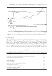
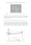
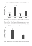
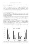
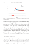
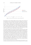
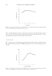
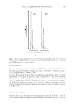
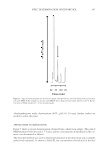
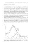
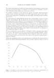
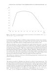
TOPICAL DELIVERY OF ANTI-INFLAMMATORY COMPOUNDS 345 UV-1700 Pharma Spec Shimadzu, Milan, Italy). Calibration curves for validated UV as- says of SG were performed on fi ve solutions in the concentration range 8—80 mg/ml. The correlation coeffi cient was greater than 0.990. Each point represented the average of three measurements, and the error was calculated as standard deviation (±SD). SG incorporation effi ciency was expressed as active recovery and calculated using equation (1): Mass of active in nanoparticles Drug recovery % 100 Mass of active fed to the system = × (1) Possible lipid interferences during UV determination of SG were also investigated by comparing the standard curve of the substance alone and in the presence of lipids. The differences observed between the standard curves were within the experimental error, thus inferring that no lipid interference occurred (data not shown). IN VITRO STUDIES Skin membrane preparation. Samples of adult human skin (mean age 36 ± 8 years) were obtained from breast reduction operations. Subcutaneous fat was carefully trimmed and the skin was immersed in distilled water at 60 ± 1°C for 2 min (16), after which SCE was removed from the dermis using a dull scalpel blade. Epidermal membranes were dried in a desiccator at approximately 25% relative humidity. The dried samples were wrapped in aluminum foil and stored at 4 ± 1°C until use. Previous research work showed the maintenance of SC barrier characteristics after storage in the re- ported conditions (17). Besides, preliminary experiments were carried out to assess the bar- rier integrity of SCE samples by measuring the in vitro permeability of [3H] water through the membranes using the Franz cell method described below. The value of calculated per- meability coeffi cient (Pm) for [3H] water agreed well with those reported earlier (18). In vitro skin permeation experiments. Samples of dried SCE were rehydrated by immersion in distilled water at room temperature for 1 h before being mounted in Franz-type diffusion cells supplied by LGA (Berkeley, CA). The exposed skin surface area was 0.75 cm2 and the receiver compartment volume was 4.5 ml. The receptor compartment was fi lled with a water–ethanol solution (50:50) (to allow the establishment of sink conditions and to sustain permeant solubilization), stirred at 500 rpm, and thermostated at 32 ± 1°C during all experiments (19,20). Approximately 100 mg of each formulation (A–H, SLN-IN, and SLN-OUT) was placed on the skin surface in the donor compartment and the latter was covered with Parafi lm® (Pechiney Plastic Packaging Company, Chicago, IL). Each experiment was run in duplicate for 24 h using three different donors (n = 6). At predetermined in- tervals, samples (200 μL) of receiving solution were withdrawn and replaced with fresh solution. The samples were analyzed for SG and DG content by HPLC as de- scribed below. The results were expressed as cumulative amount of SG and DG per- meating the SCE membranes after 24 h. Statistical analysis of data was performed using student’s t-test.
Purchased for the exclusive use of nofirst nolast (unknown)
From: SCC Media Library & Resource Center (library.scconline.org)