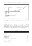
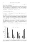
JOURNAL OF COSMETIC SCIENCE 346 IN VIVO STUDIES To investigate the relationship between in vitro skin permeation data and in vivo topical anti-infl ammatory activity, we evaluated the ability of the formulations that showed the best in vitro profi le, to inhibit the UV-induced skin erythema on healthy human volunteers. Volunteers recruitment. In vivo experiments were performed on a group of ten volunteers of both sexes in the age range 25–35 years. They were recruited after medical screening, including the fi lling of a health questionnaire, followed by physical examination of the application sites. After they were fully informed on the nature of the study and on the procedures involved, they gave their written consent. The participants did not suffer from any ailment and were not on any medication at the time of the study. They were rested for 15 min prior to the experiments and room conditions were set at 22±2°C and 40– 50% relative humidity. In vivo anti-infl ammatory activity. UVB-induced skin erythema was monitored by using a refl ectance visible spectrophotometer X-Rite model 968 (X Rite Inc., Grandville, MI), calibrated and controlled as reported earlier (12,21). Refl ectance spectra were obtained over the wavelength range 400–700 nm using illumi- nant C and 2° standard observer. From the spectral data obtained, the erythema index (EI) was calculated using equation (2) (22): ª § · § ·º = + + - + « ¨ ¨ ¸» « © ¹ © ¹» 560 540 580 510 610 1 1 1 1 1 EI 100 log 1.5 log log 2 log R R R R R (2) where 1/R is the inverse refl ectance at a specifi c wavelength (560, 540, 580, 510, and 610). The skin erythema was induced by UVB irradiation using a UVM-57 ultraviolet lamp (UVP, San Gabriel, CA) whose specifi c parameters are reported elsewhere (12). The minimal erythemal dose (MED) was preliminarily determined, and an irradiation dose corresponding to twice the value of MED was used throughout the study. For each subject, seven sites on the ventral surface of each forearm were defi ned using a circular template (1 cm2) and demarcated with permanent ink. One of the seven sites of each forearm was used as control, three sites were treated with 300 mg of formulation D, and the remaining three with 300 mg of formulation G. The preparations were spread uni- formly by means of a solid glass rod and then the sites were occluded for 6 h using Hill Top Chambers (Hill Top Research, Cincinnati, OH). After the occlusion period, the chambers were removed and the skin surfaces were gently washed to remove the gel and allowed to dry for 15 min. Each pretreated site was exposed to UVB irradiation 1, 3, and 6 h (t = 1, t = 3 and t = 6, respectively) after gel removal and the induced erythema was monitored for 52 h. EI baseline values were taken at each designated site before applica- tion of gel formulation and they were subtracted from the EI values obtained after UVB irradiation at each time point to obtain ΔEI values. For each site, the area under a curve (AUC) was computed using the trapezoidal rule. The volunteers were again recruited to complete the experimentation after a washout period of 2 weeks and the same experimen- tal procedure was repeated for the formulations SLN-IN and SLN-OUT.

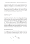
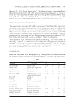
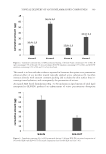
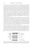
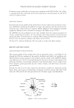
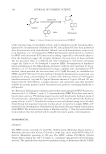
TOPICAL DELIVERY OF ANTI-INFLAMMATORY COMPOUNDS 347 To better outline the results obtained, the PIE was calculated from the AUC values using equation (3): (C) (T) (C) AUC - AUC Inhibition (%) 100 AUC = × (3) where AUC(C) is the area under the response/time curve of the vehicle-treated site (con- trol) and AUC(T) is the area under the response/time curve of the drug-treated site. Statis- tical differences of in vivo data were determined using repeated measure analysis of variance (ANOVA) followed by the Bonferroni–Dunn post hoc pairwise comparison pro- cedure. A probability, p, of less than 0.05 was considered signifi cant in this study. HPLC ANALYSES DG quantifi cation was effected by HPLC. The HPLC apparatus consisted of a Shimadzu LC-10 AT VP (equipped with a 20-μl loop injector and a SPD-M10A VP Shimadzu pho- todiode array UV detector. Chromatography was performed using a Symmetry Shield C18 RP column (particle size, 5 μm, 250 × 4.6 mm i.d.. Phenomenex, Torrance, CA). The mobile phase was composed of 30% water (pH 3 adjusted with phosphoric acid) and 70% acetonitrile and the detection was effected at 250 nm. The fl ow rate was set at 1 ml/ min. The retention time was 7.5 min. UV ANALYSES SG quantifi cation was effected by UV analyses. The UV apparatus consisted of a spectro- photometer UV-1700 PharmaSpec, Shimadzu. Calibration curves for validated UV assays of SG were performed on fi ve solutions in the concentration range 8—80 mg/ml using a wavelength of 240 nm. The correlation coeffi cient was greater than 0.990. Each point represented the average of three measurements, and the error was calculated as standard deviation (±SD). RESULTS AND DISCUSSION PREPARATION AND CHARACTERIZATION OF SG-LOADED SLN SG-loaded SLN were prepared with Compritol® 888 ATO (glyceryl behenate, tribe- henin), a mixture of mono-, di-, and triglycerides of behenic acid (C22) as the solid lipid and Pluronic® F68 (poloxamer 188) as the surfactant. We decided to use these ingredients after a lipid screening for the identifi cation of matrices for SG incorpora- tion, which pointed out a high affi nity of the active compounds toward Compritol® 888 ATO (data not shown). The use of SLN strategy to optimize DG permeation profi le resulted to be diffi cult, because the active ingredient, even if showed a chem- ical structure similar to SG, was very hydrophilic and consequently unsuitable to be formulated in a SLN system.
Purchased for the exclusive use of nofirst nolast (unknown)
From: SCC Media Library & Resource Center (library.scconline.org)