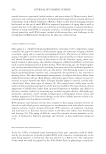
INHIBITION OF MELANIN CONTENT BY PUNICALAGINS 447 All solvents were HPLC grade from Fisher Scientifi c (Pittsburgh, PA). Cell culture media and reagents were from Life Technologies (Grand Island, NY), and all other chemicals were obtained from Sigma-Aldrich (St. Louis, MO). PREPARATION OF POMEGRANATE EXTRACT Fresh pomegranate fruit (Punica granatum) was harvested from our Nutrilite Farm (Lake- view, CA) in July 2008. The fruit (including peels) were macerated and extracted for 2 h at 60°C in a 75–80% (w/w) ethanol solution using a ratio of 1:4 (pomegranate : solvent). The extract was pressed, fi ltered, and the ethanol removed under vacuum. The aqueous extract was spray dried without carriers and stored under ambient conditions until used. The over- all yield of extract was approximately 10%. The total punicalagins content and structure was confi rmed by HPLC–MS, as reported earlier by our laboratory (17). CELL CULTURE Melan-a cells were purchased from Welcome Trust Functional Genomic Cell Bank, St. George Medical School, University of London, London (United Kingdom). Cells were grown in a 37°C, 10% CO2 incubator in RPMI-1640 media supplemented with 10% fetal bovine serum, 1% penicillin/streptomycin, 1% amphotericin B, 2 mM L -glutamine, and 200 nM tetradecanoyl phorbol acetate (TPA). Melan-a cells at passage 30 or less were used for the experiments (18). MELANIN INHIBITION ASSAY Melan-a cells were seeded at a density of 5 × 10⁴ cells per well in 24-well tissue culture plates and grown overnight. The cells were treated, in triplicate, with the indicated concentrations of pomegranate extract. Phenylthiourea (60 μg/ml), a known tyrosinase inhibitor, was used as a positive control. All compounds were prepared in 70% dimethyl sulfoxide (DMSO) and diluted to respective concentrations with cell culture medium. Melan-a cells were treated with compound for 4 days, refreshing the compound and supple- mented media at the end of day 2. Following treatment, melanin was extracted from the cells as described by Ni-Komatsu. Briefl y, the cell media were removed and the cells were lysed. The lysed cells were centrifuged, and the resulting pellet was washed with ethanol : ether (1:1) solution and then solubilized in 100 μl of 20% DMSO in 2 N NaOH. The melanin extract was transferred to a 96-well, clear-bottom plate, and total melanin content determined by reading absorbance at 490 nm on a SpectraMax M5 microplate reader (Molecular Devices, Sunnyvale, CA). All the samples were normalized to total protein content and reported as percent of melanin in untreated controls (19,21). The protein content in each sample was de- termined using the BCA protein assay kit (Pierce, Rockford, IL) as outlined by the manufac- turer. An identical set of treated Melan-a cells were used to determine cell viability. CELL VIABILITY ASSAY Cell viability was determined, following treatment of Melan-a cells with pomegranate fruit extracts, using WST-1 assay reagent (Pierce, Rockford, IL) as outlined by the manufacturer.

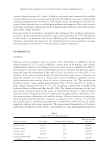
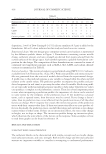
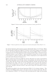
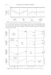
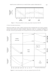
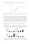
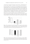
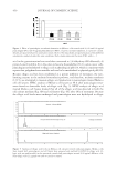
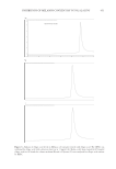
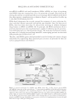
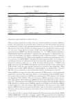
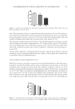
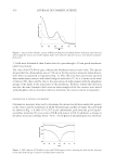
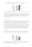
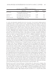
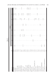
JOURNAL OF COSMETIC SCIENCE 448 Briefl y, the cell media were replaced with phenol red-free RPMI-1640 media, WST-1 reagent was added, and the cells incubated for 4 h in a 37°C, 10% CO2 incubator. The plate was read at 480 nm using a SpectraMax M5 microplate reader and cell viability was determined by measuring production of formazan dye by metabolically active cells. The results were analyzed and reported as percent of untreated controls. HPLC ANALYSIS OF ELLAGIC ACID IN CELL MEDIA AND LYSATES Melan-a cells treated with punicalagins as described earlier for the melanin inhibition assay and Melan-a cells treated for 48 h with 50 μg/ml ellagic acid were prepared for HPLC analysis of ellagic acid content. Cell media were subsampled and the cells lysed as described earlier. HPLC fractionation was achieved using an Agilent Technologies, Santa Clara, CA HP1100 System equipped with photodiode-array detection and a Waters 4 μm NovaPak column (250 × 4.6 mm). Samples were separated with a 0.2% phosphoric acid [v/v with deionized (DI) water] and acetonitrile (ACN) elution gradient as outlined in Table I. The column elution rate was held at 1 ml/min at ambient temperature with an injection volume of 10 μl for both samples and standards. The chromatogram was taken at 252 nm, integrated, and analyzed with Agilent Chemstation, a chromatography data system. An ellagic acid standard curve from 4 to 400 μg/ml (r2 0.99) was used for this analysis. Ellagic acid standards were used as positive controls with a lower detectable limit of 11 μg/ml. RESULTS AND DISCUSSION To investigate the effect of pomegranate fruit extract on melanin production, we used an in vitro Melan-a melanocyte cell culture model. Melan-a melanocytes are derived from normal mouse embryonic skin they are pigmented and dendritic, and retain almost all the characteristics of primary cells other than phenotypic variability and senescence. The cells have been shown to be an excellent model for screening antimelanogenic inhibitors for basal skin lightening products (19). Treatment of Melan-a cells with pomegranate fruit extract, standardized to 20% punicalagins, resulted in inhibition of total melanin production. Melanin content was reduced by approximately 40% and 60% at extract Table I HPLC Column Elution Gradient for Ellagic Acid Gradient Mobile Phase Time (Minutes) 0.2% Phosphoric Acid (v/v DI Water) (%) Acetonitrile (%) 0.0 92 8 12.0 90 10 14.0 88 12 26.0 78 22 29.0 78 22 29.5 92 8 32.0 92 8
Purchased for the exclusive use of nofirst nolast (unknown)
From: SCC Media Library & Resource Center (library.scconline.org)