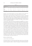
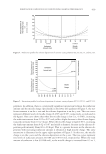
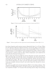
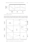
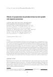
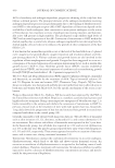
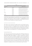
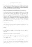
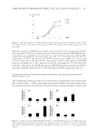
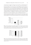
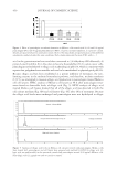
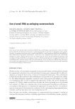
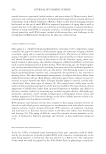
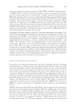
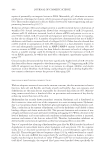
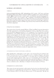
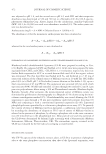
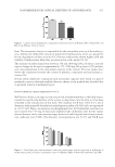
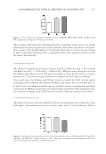
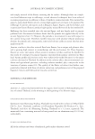
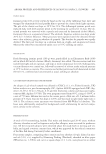
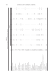
NANOBERRIES FOR TOPICAL DELIVERY OF ANTIOXIDANTS 475 form. The monomeric fraction is responsible for the antioxidant activity of the anthocya- nins, whereas the deductible amount of polymerized anthocyanins serves as a parameter for the degradation of their activity (42). Previous studies have shown higher yield and stability of anthocyanins when they are extracted in acidic media (53,54). The amounts of anthocyanins were between 350 and 1000 mg/100 g of extract, into the expected range for the species (approximately 175–3500 mg/100 g extract) (55) and they were also proportional to the antioxidant capacity in the extracts. Previous studies have shown the correlation between the content of phenolic compounds and antioxidant ac- tivities (56). Similar yields of phenolic compounds (and antioxidant capacity) were found in a pool of methanolic extracts obtained parallely (data not shown, as the method was discarded due to potential toxicity of methanol traces). PHYSICOCHEMICAL CHARACTERIZATION OF UL-B NaChol was chosen as an edge activator to provide ultradeformability to the lipid matrix, toward the need for the delivery of the actives to deep layers into the skin as it has been remarked at the introduction of this work. The resultant UL-B were 100.6 ± 0.1 nm of diameter, with unimodal distribution (polydispersion index of 0.042) and a zeta potential of −10.57 mV. Their concentration in phospholipids was 46.85 μmol/ml, and their ex- tract content was 7.93 mg of extract per milliliter (the calibration curve for the extract showed linearity in the concentration range between 0 and 6 mg/ml with a linear correla- tion coeffi cient over 0.999). The effi ciency of encapsulation was 52.5% and UL-B were Figure 1. Content of total polyphenolic compounds in ethanolic extracts of Millenia (ME), O’Neal (OE), and Blue Crisp (BE) per 100 g of extract. Figure 2. Total (light gray) and monomeric (dark gray) anthocyanin content (expressed as milligrams of anthocyanin in 100 g of extract) in ethanolic extracts of Millenia (ME), O’Neal (OE), and Blue Crisp (BE).

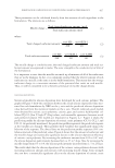
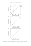
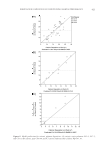
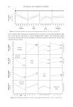
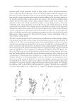
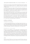
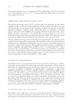
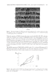
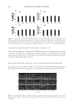
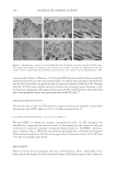
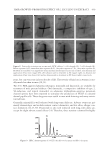
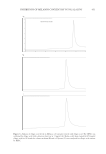
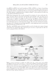
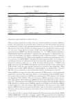
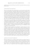
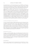
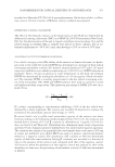
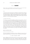
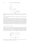
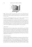
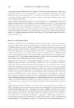
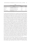
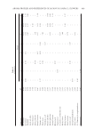
JOURNAL OF COSMETIC SCIENCE 476 3.5-fold more deformable than L when forced to pass through a 50-nm pored membrane under low pressure. The color of the UL-B was gray, whereas the blueberry extracts were violet. The spectra showed that the chromophore seen at 520 nm in the free extracts showed a bathochromic shift when incorporated to liposomes (Fig. 3). This effect has been previously reported when anthocyanins interact with other biological molecules (57). As it is shown in the work of Atrooz (58), this could be due to the association of anthocyanins with the phosphate groups of the lipids in the inner layer of the membrane. Evidence toward this direction was that the same chromatic shift was seen when samples of the free extracts were mixed with phosphate salts as described earlier to determine the extract content in liposomes. DIFFERENTIAL SCANNING CALORIMETRY Calorimetric measures were used to determine the interaction between molecules present in the extract and the membrane of UL-B. Thermotropic profi les of empty UL and UL-B are shown in Fig. 4. A shift of +0.25°C in the endothermic peak from the gel to liquid– crystalline transition (Tm) was seen in UL-B with respect to UL. In addition, a change in the phase transition enthalpy (from −59 to −25.6 J/μmol of phospholipids) was observed. Figure 3. Spectra of the ethanolic extract of Millenia (dark plot) and bathochromic shift (gray plot) observed after loading the extract into UL-B. Similar shifts were observed when free extracts were mixed with phos- phate salts. Figure 4. DSC analysis of UL (fi lled circles) and UL-B (empty circles), showing the shift and the lowering on the peak from the gel to liquid–crystalline phase transition.
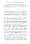
Purchased for the exclusive use of nofirst nolast (unknown)
From: SCC Media Library & Resource Center (library.scconline.org)