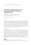
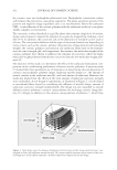
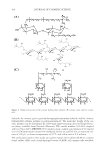
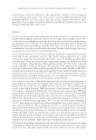
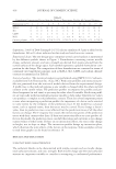
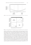
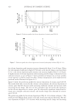
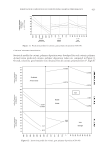
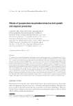
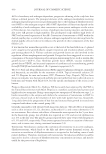
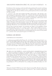
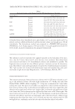
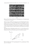
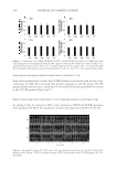
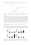
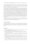
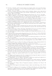
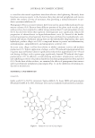
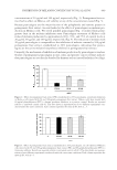
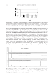
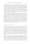
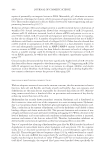
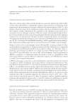
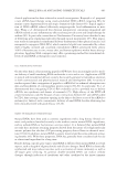
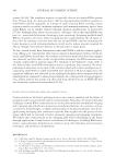
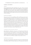
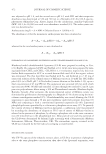
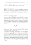
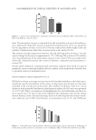
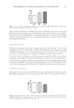
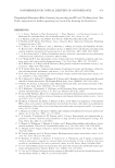
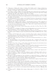
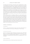
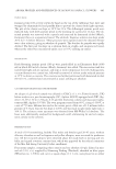
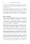
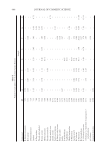
INHIBITION OF MELANIN CONTENT BY PUNICALAGINS 449 concentrations of 50 μg/ml and 100 μg/ml, respectively (Fig. 2). Pomegranate fruit ex- tract had no effect on Melan-a cell viability at any of the concentrations tested (Fig. 3). Because punicalagins are the major fraction of the polyphenols and tannins present in pomegranate fruit extract, we next looked at the effects of punicalagins on melanin pro- duction in Melan-a cells. We tested purifi ed punicalagins (Fig. 1) isolated from pome- granate fruit in the melanin inhibition assay. Punicalagins treatment of Melan-a cells reduced melanin production by approximately 60%, 70%, and 75% of control levels at 20 μg/ml, 60 μg/ml, and 100 μg/ml, respectively (Fig. 4). The reduction of melanin with 20 μg/ml punicalagins is comparable to the inhibition of melanin content by 100 μg/ml pomegranate fruit extract, standardized to 20% punicalagins, indicating that punica- lagins are the active melanin biosynthesis inhibitor in pomegranate fruit extract. Currently, the mechanism of inhibition of melanin production by punicalagins is unclear. Studies of pomegranate punicalagins in oral, colon, and prostate cancer cell lines indicate that punicalagins are not directly absorbed by humans and are instead hydrolyzed to ellagic Figure 2. Effect of pomegranate fruit extract (PE), standardized to 20% punicalagins, on melanin formation in Melan-a cells treated with 50 and 100 μg/ml pomegranate fruit extract. Melan-a cells were treated with 60 μg/ml phenylthiourea (PTU), a known tyrosinase inhibitor, as a positive control. Results are reported relative to untreated control cells (C). The data shown is representation of two different experiments con- ducted under the same conditions. Each column represents the mean ± SD, with n = 3. Figure 3. Effect of pomegranate fruit extract, standardized to 20% punicalagins, on cell viability of Melan-a cells treated with 50 and 100 μg/ml pomegranate fruit extract (PE), and 60 μg/ml phenylthiourea (PTU), a tyrosinase inhibitor. Results are reported relative to untreated control cells (C). The data shown are represen- tations of two different experiments conducted under the exact same conditions. Each column represents the mean ± SD, with n = 3.

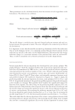
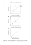
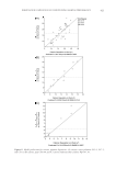
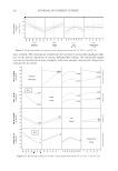
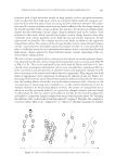
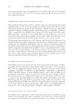
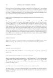
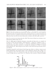
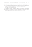
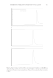
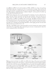
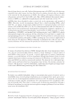
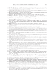
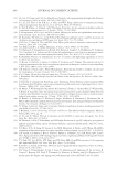
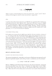
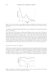
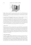
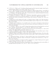
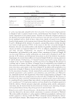
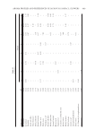
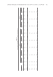
JOURNAL OF COSMETIC SCIENCE 450 acid in the gastrointestinal tract and then converted to 3,8-dihydroxy-6H-dibenzo(b, d) pyran-6-one (Urolithin A) in the colon to become bioavailable (20). In colon cancer cells, punicalagins are hydrolyzed to ellagic acid at physiological pH (10), which is consistent with reports that polyphenols are unstable and tend to be metabolized at physiological pH (16). Because ellagic acid has been established as a potent inhibitor of tyrosinase, the rate- limiting enzyme in the melanin biosynthesis pathway, and therefore, melanin synthesis (12,15), we attempted to measure ellagic acid production in punicalagins treated Melan-a cell cell lysates. HPLC analysis of Melan-a cell lysates at 48 h after punicalagins treat- ment found no detectable levels of ellagic acid (Fig. 5). HPLC analysis of ellagic acid treated Melan-a cell lysates showed that all of the ellagic acid was detected in both the cell culture medium (Fig. 6B) and cell lysates (Fig. 6C) after 48 h of treatment. Because the ellagic acid levels were unchanged and punicalagins were not hydrolyzed to ellagic Figure 4. Effect of punicalagins on melanin formation in Melan-a cells treated with 20, 60 and 100 μg/ml punicalagins (PG), and 60 μg/ml phenylthiourea (PTU), a known tyrosinase inhibitor, as a positive control. Results are reported relative to untreated control cells (C). The data shown are representations of two different experiments conducted under the same conditions. Each column represents the mean ± SD, with n = 3. Figure 5. Analysis of ellagic acid levels in Melan-a cell cultures treated with punicalagins. Melan-a cells were treated with punicalagins, and cell lysates were prepared and analyzed by HPLC for ellagic acid. El- lagic acid was undetectable in the untreated controls and punicalagins treated (100 μg/ml) Melan-a cells. Pure ellagic acid (11 μg/ml) was used as a positive control.
Purchased for the exclusive use of nofirst nolast (unknown)
From: SCC Media Library & Resource Center (library.scconline.org)