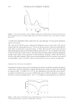
NANOBERRIES FOR TOPICAL DELIVERY OF ANTIOXIDANTS 473 recorded in a Shimadzu UV–Vis 160-A spectrophotometer. On the basis of this, a calibra- tion curve at 524 nm of extract of Millenia variety in ethanol was prepared. DIFFERENTIAL SCANNING CALORIMETRY The effect of the ethanolic extracts on the lipid matrix of the UL-B was determined by differential scanning calorimetry (DSC) on a MDSC Q-200 TA Instruments (New Castle, DE). For the determination of the (gel to liquid–crystalline) transition phase (Tm) and the related change in enthalpy (ΔHcal), samples were placed in T-zero capsules, fi rst in iso- thermal equilibrium at −40°C for 5 min, then heating to 20°C at a rate of 10°C/min. ANTIOXIDANT ACTIVITY DETERMINATION METHODS Free radical scavenging activityThe ability of the extracts to donate electrons or a hydro- gen atom to the stable free radical DPPH was determined as a measure of their radical scavenging antioxidant activity (48). In brief, aliquots between 0.057 and 0.113 mg of extract per milliliter were added by triplicating to a 0.0031% (w/v) DPPH solution in methanol. After a 30-min incubation at room temperature in the dark, the remnant DPPH was determined by reading the absorbance at 515 nm against a blank of metha- nol. The remnant DPPH is inversely proportional to the free radical scavenging activ- ity of the antioxidant (49). BHT and DPPH solution were used as a positive control and inhibition blank, respectively. The inhibition percentage of DPPH (I%) was calcu- lated (50) as: blank sample blank A A¯ I% A ¡ ° q100 ¡ ° ¢ ± IC50 values, corresponding to concentrations inhibiting a 50% of the free radical were obtained by a linear regression. The activity was recorded 30 days later to evaluate the retention of the antioxidant capacity after storage at −18°C. β-carotene–linoleic acid assayThe total antioxidant capacity of the extracts was deter- mined according to the following method adapted from (39,51,52). An emulsion was formed from a mixture of 0.5 ml β -carotene in chloroform at 1 mg/ml 0.02 ml lin- oleic acid and 0.2 ml Tween 80. Chloroform was eliminated under rotary evaporation and then 50 ml of oxygen-saturated milliQ water was added under vigorous agitation. The emulsion was aliquoted in eppendorf tubes and samples from each extract (0.85 mg of extract per milliliter) were added. BHT was used as a positive control and absolute ethanol as a negative control. The absorbance of β -carotene at 460 nm determined against a blank (the same emulsion lacking β -carotene) was recorded as a function of time in the presence of the extracts and compared with that of the negative control. 42 h later (t), when the absorbance of the negative control was reduced to 10% of the absorbance at time zero, the percentage of antioxidant activity (% AA) was determined as follows:

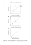
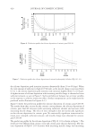
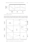
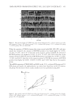
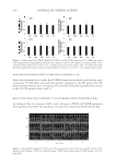
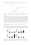
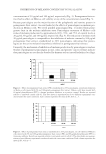
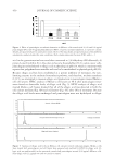
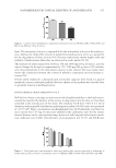
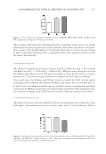
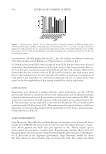
JOURNAL OF COSMETIC SCIENCE 474 0 0 0 0 1 ( % 100 t t A A¯) AA A A ¡ ° ¡ ° ¢ ± where A0 and At are the absorbances at time zero and at time t, and A00 and At0 the ab- sorbances of the negative control at time zero and time t, respectively. CELLS A cell line derived from human keratinocytes (HaCaT) was supplied by Dr. E. Salvatierra of Fundación Instituto Leloir (Buenos Aires, Argentina). Cells were cultured in MEM (Gibco, Life Technologies, Carlsbad, CA) supplemented with 10% fetal calf serum (FCS), 1% antibiotic/antimycotic (PAA Laboratories GmbH, Pasching, Austria) (penicillin 10,000 U/ml, streptomycin sulphate 10 mg/ml, amphotericin B 25 μg/ml) and 2 mM glutamine, at 37°C in 5% CO2 and 95% humidity. CYTOTOXICITY IN HaCaT CELLS Cell viability was measured by the MTT assay. HaCaT cells were seeded at a density of 3 × 10⁴ cells per well onto 96-well fl at-bottom plates and grown for 24 h at 37°C. Then, the medium was replaced by 100 μl of fresh MEM with 5% FCS containing dilu- tions of the ethanolic extracts of each variety (3, 30, and 300 mg of fresh fruit per mil- liliter), UL-B suspension (2 and 14 mg of fresh fruit per milliliter), empty UL (at the same concentration in lipids than in UL-B) and untreated control. Cells were incubated at 37°C for 24 h. After incubation, 110 μl of 0.45 mg/ml MTT were added to cells at- tached to plates. After 3 h of incubation, MTT solution was removed, the insoluble formazan crystals dissolved with 100 μl of dimethylsulfoxide , and absorbance was measured at 570 nm using a microplate reader (Dynex Technologies, MRX tc, Chantilly, VA). Viability of cells was expressed as a percentage of the viability of cells grown in the medium. RESULTS AND DISCUSSION PHENOLICS AND ANTHOCYANIN CONTENT The amount of polyphenolic compounds in each variety of blueberries was expressed as mil- ligrams of equivalents of gallic acid in 100 g of extract. A calibration curve was obtained: 2 0.004 0.0064 ( 0.9999) y x R Extracts of Millenia and O’Neal showed nearly twice the amount of phenolic compounds than that of the Blue Crisp extract (Fig. 1). Similarly, the anthocyanins content of Mil- lenia and O’Neal extract were more than twice the amount in Blue Crisp, as presented in Fig. 2. More than 90% of the anthocyanins in all extracts were found in their monomeric
Purchased for the exclusive use of nofirst nolast (unknown)
From: SCC Media Library & Resource Center (library.scconline.org)