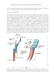
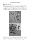
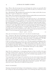
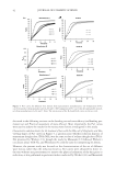
NEW ASPECTS OF THE STRUCTURE OF HUMAN HAIR 23 The high tolerance of the wall to the severe chemical treatment may be attributed to the proteins that were heavily cross-linked with the isopeptide [ε-γ(glutamyl)lysine] and disulfi de bonds (3–6 and 15–20 bonds per 1000 amino acid residues, respectively) (29, 30). On the other hand, the Cu was found to take the trowel-like shape, consisting of a trans- parent CuB and a CuH Figure 1(B) and the inset, cf. Diagram 1 (position: 10-m). The spatial arrangement of CuB and CuH were judged in Figure 7, i.e., CuH was attached to the outer surface of CuB cf. the schematic illustration in Diagram 2 (position: 6-b and 5-i). The cell itself, however, was not so stable on the mechanical impact given in the Figure 4. (A) A block of the cortical cells PL20× objective bars 25 μm. Hair I was heated in an aqueous mixture of 6.4 M urea, 3.2 wt.% SDS and 20 wt.% ME at 80°C for 7 h, followed by subjecting to the cutting process II CBB-staining. (B) Plenty of the fi brous substance in the cortical cells’ block bar 50 μm. The specimen was prepared by treating hair III in a manner similar to that mentioned in panel A, except for Gentian violet staining. Figure 5. (A) Cortical cell (Co), the blade-like shaped (CuB), and handle-like shaped (CuH) parts of the cuticular cell bar 50 μm cf. Figure 1. (B) CuB overlapped the other to display a tile roof-like or so-called “scale” pattern bar 50 μm. Every CuB possessed a nucleus (N) with 3–7 small black nucleoli. The specimens of panels A and B were obtained by heating hair (III) in a solution of 8 M urea and 4 wt.% SDS (without using ME) at 85°C for 15 h, followed by subjecting to the cutting process I Gentian violet staining.

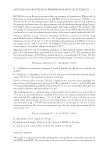
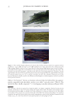
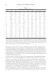
JOURNAL OF COSMETIC SCIENCE 24 cutting process that it was split into CuB and CuH, and CuB was further broken into CuB’ Figure 1(A). CuB’ was therefore recognizable as a small fl at substance having at least one jagged edge, being very similar in shape to the traditional Cu (2,9) cf. Diagram 2 (position: 3-i). On the other hand, CuH resembled the Co not only in shape but also in the richness of the fi brous substances (Mf) though the former part appeared to be more Table I Amino Acid Compositions of the Hollow Fibers and Whole Cuticle Amino acid Residues per 1000 amino acid residues Hollow fi ber from Hair III Whole cuticlea Cysteine + half Cysteine 155 180.8 Aspartic acid 54 32.4 Threonine 47 46.1 Serine 133 160.7 Glutamic acid 90 89.2 Proline 97 105.4 Glycine 93 88.4 Alanine 54 54.1 Valine 63 72.7 Methionine 1 4.6 Isoleucine 27 22.3 Leucine 52 44.6 Tyrosine 25 21.1 Phenylalanine 14 11.7 Lysine 39 34.4 Histidine 5 5.2 Arginine 36 26.4 aCited from reference 7. Figure 6. (A) The inner substances were squeezed out from hair (I) when the fi ber was heated in an aqueous solution of 6.4 M urea, 3.2 wt.% SDS and 20 wt.% ME at 80°C for 2 h staining with the Giemsa’s solution PL20× objective bar 50 μm. (B) The hollow fi ber PL20× objective bar 50 μm. Hair III was heated in 7 M urea, 3.5 wt.% SDS and 15 wt.% ME at 80°C for 7 h and subjected to the cutting process II silver staining (C) The cross section of the hollow fi ber bar 50 μm. Hair IV was heated in 7.6 M urea, 3.8 wt.% SDS and 5 wt.% ME at 80°C for 7 h, then subjected to the cutting process III Gentian violet staining. The wall is mainly consisted of the blade-like shaped parts (CuB) of the cuticular cells. The black amorphous substances on the inner surface of the wall are most likely the remnant of the handle-like shaped part (CuH) cf. Figure 9(B). The inset is the enlarged view of a part of the wall the small black spots may be melanin particles bar 5 μm.
Purchased for the exclusive use of nofirst nolast (unknown)
From: SCC Media Library & Resource Center (library.scconline.org)