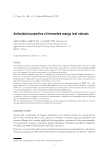
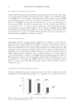
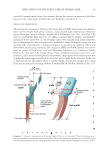
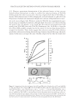
PRACTICAL SELECTING METHOD OF WAVE LOTION FOR HAIR DRESSER 37 form is remained. In our method, a single hair was used and the bending set was mea- sured using the devised apparatus as follows. In the bending set experiment, a single hair fi ber was placed on the bending apparatus in the manner illustrated in Figure 3. The fi ber was equipped with small weights at both ends and was wound at the root of a 10-mm-diameter cylinder on the device shown in the fi gure and vertically held in water at 30°C for 5 min. While holding the fi ber in water, the four nuts were evenly tightened to fi x the fi ber onto the device. The weights were then removed and the water on the surface of the device was wiped off. The device fi xing the wet fi ber was then placed into a 100-ml beaker containing 50 ml of a given waving lotion for t min, and subsequently the device was treated in the pre- pared beaker under the same conditions as those described in Steps 3, 4, and 5 as shown in Figure 2. When the treatment was fi nished, the fi ber was cut at the point of intersection, separated from the device, and immediately transferred into a Petri dish fi lled with water at 30°C. The Petri dish was then placed on a photocopy machine. By covering the fi ber within the Petri dish with a white PVC disk, a good quality photocopy could be obtained. The pho- tocopy of each bending treated fi ber was in the shape of an arc, and the radius of the arc Figure 3. Device used for the permanent treatment of the bending set. A single wet hair wound at the root of the cylinder and fi xed using silicon plates that are tightened to the screws by four bolts.

JOURNAL OF COSMETIC SCIENCE 38 was measured from a copy enlarged by 100%. The extent of the bend induced by the bending treatment, Sℓ (bend) (%), was calculated as follows: Sℓ(bend) = (5.0 ∕ r∕2) × 100%, (3) where r is the bend radius of the fi ber derived from the doubly enlarged copy. WAVE EFFICACY IN A BEAUTY PARLOR Practical treatment measurements were performed by a profi cient hairdresser, and the results were reported to us. A small tissue wound on a rod was treated, and the resulting assessment, referred to as the “wave effi cacy,” was calculated using the spiral tissue length, length of the wave and diameter of the rod: wave effi cacy = 100 × N × D/ℓ, (4) where l is the length of the spiral tissue, N is the wave length, and D is the diameter of the rod. MEASUREMENT OF THE 20% INDEX The path connecting the water contained in columns A and B was opened by operating the three-way stopcock, and both columns were fi lled with water in order to maintain a constant water level in Column A. The tensile tester was programmed to alternately load a 20% strain and then unload to the original position at a rate of 5 mm/min. The result- ing hysteresis curve provided the 20% index. MICROSCOPY OF CROSS SECTIONS OF THIOGLYCOLATE-TREATED HAIRS White hair fi bers were cleaned with a 1 g/100 g lauryl sulfate aqueous solution, washed with water, immersed for 3 min, wiped with paper, followed by immersion in the respective reducing agent (0.6 M, pH 8.6) for 5 min. The reduced fi bers were washed with deashed water for 3 min and subsequently dried at room temperature. Thioglycolate-treated hairs were cryoprotected in a graded series of sucrose (5, 10, and 20% in a 0.1 M phosphate buffer, pH 7.3) on ice and embedded in optimal cutting tem- perature compound (Sakura Finetechnical Co. Ltd., Tokyo, Japan) before freezing on a metal block prechilled with liquid nitrogen. Frozen hairs were cross sectioned in 10-μm thick sections in a Cryostat (Leika CM 1850, Nussloch, Germany), air-dried on micro- scopic slides, and post-fi xed in 4% paraformaldehyde dissolved in a 0.1 M phosphate buffer (pH 7.3) for 5 min. The sections were prewashed in a 0.1 M TrisHCl buffer at pH 9.5, stained with 0.1% methylene blue for 5 min, and then rinsed in the same buffer. Microscopic images were captured under epi illumination using a Nikon E600 optical system (Nikon Instech Co. Ltd., Tokyo, Japan) equipped with an Olympus DP70 camera (Olympus, Tokyo, Japan). The captured images were density scanned on a computer us- ing a National Institute of Health (NIH) Image analyzer with the aid of a software by Celisene Olymoas Co. Ltd (Tokyo, Japan).
Purchased for the exclusive use of nofirst nolast (unknown)
From: SCC Media Library & Resource Center (library.scconline.org)