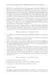
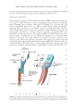
NEW ASPECTS OF THE STRUCTURE OF HUMAN HAIR 25 easily stained with Congo red than Co Figures 1(A) and 2 cf. Diagram 1 (position: 2-m vs. 8-m). By the way, honeycomb-like and overlapping scale-like patterns were observed in the inner and outer surfaces of the CuB region, respectively Figure 8 cf. Diagram 1 (position: 9-a and 9-f). A formation of these unique patterns is further discussed below. CUTICULAR THIN PLATE The present study indicated defi nitely the presence of a thin plate (CuP 0.4–0.6 μm in thickness) in the Cu region Figures 9(A), 9(B), and 10 cf. Diagram 1 (position: 9-e) and Diagram 2 (3-a and 4-g). CuP was a transparent material and its presence was revealed only by means of phase contrast microscopy conversely, CuP was neither colored by polarized light microscopy nor stained with the dyes such as CBB and Gentian violet. When the infants’ hair (I and II) were treated with the high concentrations of ME or for a long reaction time, CuP was isolated as a transparent fi lm-like substance Figure 9(C). Though the formation of CuP has not been explained well, it is conceivable that CuB fuse partially with the neighboring ones to build the honeycomb-like structure [Figure 8(A) and 8(B)], gradually merging into the single thin plate in the boundary zone between CuB and CuH. In fact, many merging sites were spotted in the inner surface of CuP see, for example, the site marked by the gray circle of Figure 10(A). By the way, CuP was extremely tolerant to heating in the aqueous solution of 20 wt.% ME. The hollow fi ber [Figure 6(B)] should not be stable without CuP in other words, the hollow structure may be decomposed if the wall were merely constituted from the overlapping Cu. It would Figure 7. The hand-like-shaped part (CuH) of the Cu was attached on the outer surface of the blade-like- shaped part (CuB) bar 40 μm. The black line-framed area is enlarged to give the inset. The CuB in these pictures appears to be somewhat short in length, presumably because a tip portion of the complete form was shaved off in the cutting process. Hair IV was heated in 8 M urea and 4 wt.% SDS (without ME) at 60°C for 6 h, followed by the cutting process III SM staining.

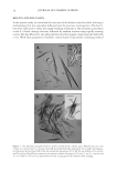
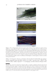
JOURNAL OF COSMETIC SCIENCE 26 also be deduced that the inner components of hair including the Co and medulla are physicochemically protected, at least to some extent, from the outside circumstance by the presence of CuP. MEDULLA This component has been considered for a long time as a loosely packed assembly of the cellular disks connected in series (1,3,31). Nevertheless, it was suggested in the present Figure 9. A cuticular thin plate (CuP) exists in between the regions of the blade-like shaped parts (CuB) and the handle-like shaped parts (CuH) of the Cu. (A) Hair III was heated in an aqueous solution of 7.4 M urea, 3.7 wt.% SDS and 8 wt.% ME at 50°C for 2 h and subjected to the cutting process II CBB staining PLL40× objective bar 50 μm. As observed in this panel, the CuB region was usually paired with the CuH region presumably because both regions were linked to each other through CuP. (B) Part of the Cu region PLL40× objective bar 50 μm. CuP is seen as a transparent substance between the CuB and CuH regions in the phase contrast microscopy. The hair III was treated as mentioned in the panel A, except for the cutting process III and double staining with CBB and Gentian violet. (C) Part of an isolated CuP PL20× bar 50 μm. Hair II was heated in an aqueous mixture of 7 M urea, 3.5 wt.% SDS and 15 wt.% ME at 80°C for 3 h and sub- jected to the cutting process III staining with Giemsa’s solution. Figure 8. (A) A honeycomb-like pattern in the inner surface of the region of the blade-like shaped parts (CuB) of the Cu PLL40× objective bar 50 μm cf. Diagram 1 (position: 9-a). (B) A so-called “scale” pattern was seen when the reverse side of the CuB region (of the panel A) was focused on note that the tiling direc- tion was opposite between the panels A and B. The specimens of both panels were prepared by heating hair IV in an aqueous mixture of 7 M urea, 3.5 wt.% SDS and 15 wt.% ME at 80°C for 7 h, followed by the cut- ting process III Gentian violet staining.
Purchased for the exclusive use of nofirst nolast (unknown)
From: SCC Media Library & Resource Center (library.scconline.org)