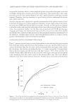
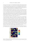
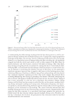
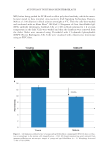
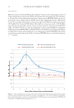
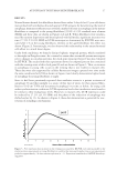
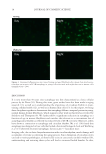
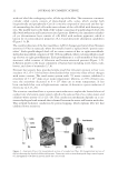
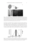
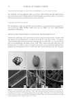
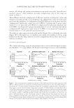
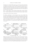
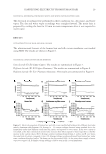
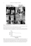
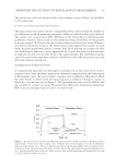
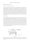
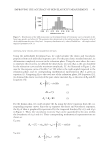
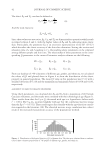
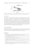
AUTOPHAGY IN HUMAN SKIN FIBROBLASTS 17 RESULTS Normal human dermal skin fi broblasts derived from either 2-day-old or 67-year-old donors were probed with antibodies directed against LC3B antigens for determining the level of autophagy. Immunocytofl uorescence revealed a marked decrease in autophagy in the mature fi broblasts as compared to the young fi broblasts [23.8% ± 1.9% standard error of mean (SEM)] and these data are shown in Figures 1A and B. When fi broblasts were synchro- nized by nutrient deprivation and then repleted with full media, signifi cant increases over time (77.9% ± 10.9% SEM) in LC3B transcripts as determined by RT-PCR were mea- sured after 2 h in the young fi broblasts, whereas in the aged fi broblasts this effect was absent (Figure 2). Interestingly, we also observed this relationship in the unsynchronized cells albeit to a much lesser degree. Under both conditions, we found a distinct biphasic temporal pattern, which correlated to the night in Zeitgeber time. As a control to ensure that our model system was respon- sive to changes in circadian patterns, the clock gene transcript for per2 was also evaluated by RT-PCR. The results from this experiment showed a temporal pattern that correlated with the evening onset of this clock gene (8) and are shown in Figure 3. Thus, an increase in autophagy in young cells occurs in the evening that is not evident in mature cells. These data were also supported by cellular fl uorescent images captured over time under the same conditions for LC3B as shown in Figure 4 and clearly demonstrate higher levels of autophagy for young fi broblasts at night. Since it had been previously reported that oxidative stress is a potent activator of autophagy (9) and that sunlight is a source of this type of stress, we then exposed fi bro- blasts to 10 J/cm2 UVA and determined autophagy over time as before. In young cells under synchronization conditions, LC3B expression levels after irradiation were found to be similar to their endogenous levels. However, in mature cells, LC3B expression could be induced by 27.2% (±5.3% SEM) and the phase of the induction of autophagy was shifted earlier by 1 h. As shown in Figure 4, these data demonstrate a potential for reac- tivation of autophagic mechanisms. Figure 3. Per2 expression that increases in the evening was assayed by RT-PCR over time in parallel to the synchronization experiments in young fi broblasts to confi rm that evening Zeitgeber time in our experiments peaked at approximately 2 h after nutrient repletion (n = 3). Expression normalized to GAPDH housekeep- ing genes.

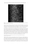
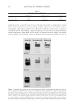
JOURNAL OF COSMETIC SCIENCE 18 DISCUSSION It is now more than 50 years since autophagy was fi rst characterized as a basic cellular process by de Duve (10). During this time, great strides have also been made in aging research (11), as well as in understanding the importance of circadian rhythm in main- taining cellular health (12), as well as in human skin cells (13). In this report, we bring these disciplines together to demonstrate that autophagy follows a temporal pattern in young, normal human dermal fi broblasts, which supports other work in this fi eld as reviewed by Sachdeva and Thompson (6). We further show a signifi cant reduction in autophagy as a function of age in mature fi broblasts and correlate this decrease to a concomitant loss of autophagosomal rhythm as refl ected by reduced levels of LC3B over time. Moreover, others have shown a connection to autophagy and circadian rhythm. Ma et al. (14) found that autophagy was disrupted in liver cells that lacked a functional biological clock and Weibking et al. (15) observed decreased autophagic factors in per1-/- knockout mice. In aging cells, due to their dissynchronization with circadian rhythm, much damage will accumulate over time accelerating the aging process. Since disruption of circadian cycles can lead to various pathologies, as observed by a rise in cancer rates in shift workers (16), understanding and restoring these cycles will lead to increased health benefi ts. In skin, Figure 4. Immunocytofl uorescence over time of young and aged fi broblasts after release from synchroniza- tion show an increase in LC3B/autophagy in young cells associated with night but not in mature cells (magnifi cation = 20×).
Purchased for the exclusive use of nofirst nolast (unknown)
From: SCC Media Library & Resource Center (library.scconline.org)