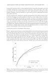
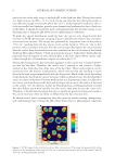
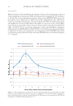
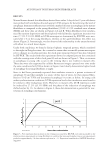
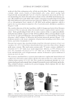
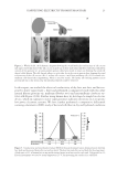
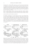
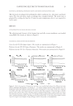
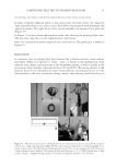
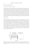
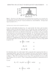
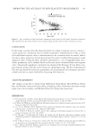
HARVESTING ELECTRICITY FROM HUMAN HAIR 25 from the buildings in the winter, where a large amount of heat energy is needed to maintain the temperature. MATERIALS AND METHODS COLLECTION OF HAIR SAMPLES AND PROCESSING Hairs from random individuals were collected, and washed with deionized water twice for an hour. This wet hair was then allowed to dry in a laminar fl ow hood at room tem- perature. One of challenging problem is to wash the hair sample and to ensure the re- moval of the ionic species (27). Thus we conducted a simple test to ensure that most of the loosely bound ionic species are removed during washing. We fi rst measured the conductivity of the deionized water to be used for washing the hair (Figure 3). Next, we kept the hair sample in this water and used a magnetic stirrer to swirl the water for 30 min, thus ensuring that all the loosely bound ionic species present in water gets dissolved in water. After the fi rst wash, we measured the conductivity of the water. We observed a signifi cant increase in conductivity, thus indicating the presence of ionic species which has leached out from the hair surface. Again we repeated the process for 30 min and measured the conductivity. We observed that the conductivity of water falls down to the baseline. This experiment ensured that the removal of the ionic spe- cies and proper washing of the hair. Figure 5. Hair bioelectric device. (A) All the components needed to fabricate a simple hair bioelectric de- vice. (B). The overall test apparatus showing the source of water vapor. (C) The bioelectric device inside the plastic casing attached to the glass tube supplying water vapor.

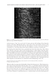
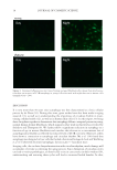
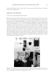
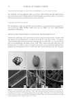
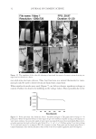
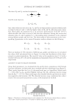
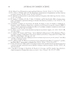
JOURNAL OF COSMETIC SCIENCE 26 COLLECTION OF MULBERRY (B. MORI) AND NON-MULBERRY (A. MYLITTA) SILK COCOONS The mulberry and non-mulberry silk cocoons were collected from the silk farmers resid- ing in the Indian states of Karnataka and Chattishgarh, respectively. These cocoons were cleaned with a dry blower and stored in a desiccator (26). SCANNING ELECTRON MICROSCOPY The morphology of the hair and silk cocoon membrane was studied using scanning elec- tron microscopy (SEM SUPRA 40VP fi eld emission SEM, Carl Zeiss NTS GmbH, Oberkochen, Germany). SELECTION OF ELECTRODE MATERIALS AND DEVELOPING THE MEASUREMENT SETUP Aluminum, platinum, and copper were chosen as the electrode metals. A glass slide was used as the holding frame, on which electrode (E1) was fi xed. Then, 150 grams of dried human hair was spread evenly covering the electrode (E1). In case of silk, the silk cocoon membrane was fi xed on top of E1. The counter electrode (E2) wire was wounded over the whole setup covering the hair/silk cocoon membrane substantially, forming the measurement device with hair/ silk sandwiched between the electrode Figure 6. Gross and scanning electron microscopic morphology of Human hair and Silk Cocoon. (A) Natu- ral human hair with black texture. (B, C) A. mylitta silk cocoon has brownish coarse texture while B. mori cocoon has a soft texture. (D) Scanning Electron Microscopy image of the surface of a human hair showing cuticle (scales) on outside of our hair. (E, F) Rough outer surface of A. mylitta with some crystals of calcium oxalates and smooth outer surface of the B. mori cocoon.
Purchased for the exclusive use of nofirst nolast (unknown)
From: SCC Media Library & Resource Center (library.scconline.org)