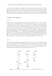
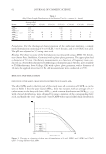
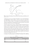
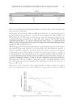
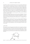
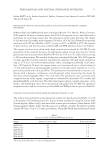
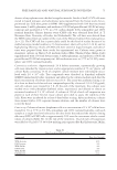
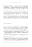
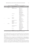
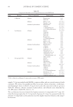
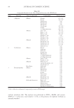
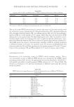
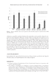
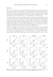
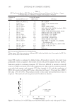
FREE RADICALS AND NATURAL SUBSTANCES IN PELOIDS 85 The use of cesium-EDTA was necessary to remove the metal ions that may interfere with the relaxation times of phospholipids. Phosphatidylcholine (PC), phosphatidylserine (PS), phosphatidylethanolamine (PE), and sphingomyelin (SM) were the main phospho- lipids in DM (in a total amount of 0.52 mg/g), while they were not detected in WM. Among the recent applications, PC shows hepatoprotective effect in oxidative-stress- mediated organic solvent liver damage (65), and it is applied in bipolar radiofrequency treat- ment of localized fat deposits (66). PS is involved in specifi c drug delivery systems (67). Table XIII reports the amount of phospholipids identifi ed in DM for sampling performed at different months in the year. As a general trend, the amount of phospholipids was relatively stable, the PC and PS being isolated in the highest amount (0.25 and 0.22 mg/g, respectively) in January, PE and SM in March (0.13 and 0.06 mg/g, respectively). ANTIOXIDANT ACTIVITY Determination of the antioxidant activity by DPPH radical scavenging analysis. The in vitro antioxidant activity of DM and WM peloids was evaluated by the DPPH radical scavenging analysis (68). In brief, the appropriate fraction was dissolved in EtOH (0.01– 100 mg/ml) and added to freshly prepared DPPH solution (6 × 10⁻⁵ M in EtOH). The decrease in absorbance (475 nm) was determined at different times until the reaction reached a plateau. Table XIII Amount of PC, PS, PE, and SM Identifi ed in DM for Sampling Performed at Different Months Compound Septembera November January March May PC 0.2 0.16 0.25 0.15 0.19 PS 0.16 0.16 0.22 0.18 0.2 PE 0.12 0.08 0.08 0.14 0.10 SM 0.04 0.04 0.03 0.06 0.03 a The amount is defi ned as milligram of phospholipid per gram of starting DM. Table XIV DPPH Radical Scavenging Properties of Polar and Nonpolar Fractions of DM and WM Peloids, Respectively Entry Sample IC50 1 DM-EtOAc 0.11 2 DM-Acetone 0.20 3 DM-MeOH 2.0 ×10⁻³ 4 DM-Heptane 95 5 DM-Cyclohexane 93 6 DM-Diisopropyl ether 90 7 WM-EtOAc 0.13 8 WM-Acetone 0.19 9 WM-MeOH 1× 10⁻³ 10 WM-Heptane 95 11 WM-Cyclohexane 93 12 WM-Diisopropyl ether 90 Each experiment was performed in triplicate.

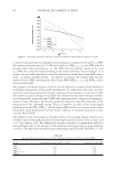
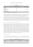
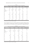
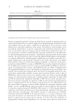
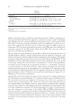
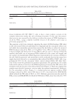
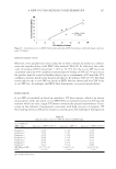
JOURNAL OF COSMETIC SCIENCE 86 The kinetic of the process was analyzed for each concentration tested, and the percentage of DPPH remaining at the steady state was estimated. This value was used to calculate the IC50 (defi ned as the concentration of substrate mg/ml that causes 50% loss of DPPH activity). Results are reported in Table XIV. The polar fractions of DM and WM showed a similar antioxidant activity (Table XIV, entries 1–3 vs. 7–9), higher than that observed for the corresponding nonpolar fractions (Table XIV, entries 1–3 vs. 4–6). In particular, EtOAc was the most active fraction in DM (IC50 2.0 × 10⁻³ mg/ml), and MeOH in WM (IC50 3.0 × 10⁻³ mg/ml). Evaluation of cytotoxic potential. The cytotoxic potential was evaluated in CHO cells only for the polar fractions, using the clonogenic assay (CA). CA enables an assessment of the differences in reproductive viability (capacity of cells to produce progeny, i.e., a single cell to form a colony of 50 or more cells) between control untreated cells and cells after treat- ment (69). Normally, only a hundred or few hundred cells are inoculated. Each viable cell grows and forms a colony. After a suitable incubation time (5–8 days), colonies are stained and counted manually. Cloning effi ciency is calculated as percentage (%) of colonies from all inoculated cells. Cytotoxicity was determined by measuring the RCE after treatment compared with cloning effi ciency of solvent control cultures. The assay was performed using a set of at least six dose levels for each extract, spaced by a factor of 2.0. DMSO was used as solvent. Results obtained indicated that after a 3-h treatment, dose level of 32.0 μg/ml proved to be toxic (RCE 20%) for all tested extracts, with the exception of WM-MeOH and DM-MeOH for which toxic levels were reached at 562.0 and 128.0 μg/ml, respectively (Tables XV–XX). Genotoxic and antioxidant activity in cultured mammalian cells. The antioxidant activity of polar and nonpolar fractions of DM and WM peloids was further evaluated in Table XV Analysis of Colony-Forming Activity for WM-Acetone Test compound Dose level (μg/ml) Mean of colonies RCE DMSO 1 317 100 WM-Acetone 1.0 311 98 2.0 310 98 4.0 276 87 8.0 200 63 16.0 171 54 32.0 10 3 Table XVI Analysis of Colony-Forming Activity for DM-Acetone Test compound Dose level (μg/ml) Mean of colonies RCE DMSO 1 298 100 DM-Acetone 1.0 292 98 2.0 265 89 4.0 224 75 8.0 143 48 16.0 54 18 32.0 12 4
Purchased for the exclusive use of nofirst nolast (unknown)
From: SCC Media Library & Resource Center (library.scconline.org)