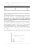
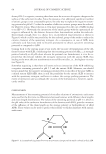
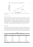
J. Cosmet. Sci., 67, 109–110 (March/April 2016) 109 Letter to the editor The Entrance Echo corresponds to Epidermis on High Frequency Skin Ultrasound XIMENA WORTSMAN, Departments of Radiology and Dermatology, Institute for Diagnostic Imaging and Research of the Skin and Soft Tissues, Clinica Servet, Faculty of Medicine, University of Chile, Las Condes, Santiago, Chile I have read with great interest the article “The top echorich band in a 50-MHz ultrasound sonogram refl ects epidermal properties” (1). I congratulate the authors for their work that demonstrates that the entrance echo, also called entry echo, (i.e., hyperechoic layer on top of the dermis) on ultrasound indeed corresponds to the epidermis. This has been an old and long discussion among the operators of high-frequency probes that has created a deep confusion in the literature. We have arrived to the same conclusion by comparing the ultrasound images of patients with psoriasis (2) that present sonographic thickening of the epidermis and patients with cutaneous ulcers that show absence of the epidermis at the site of the ulcer on ultrasound, which has been also correlated with histology (3). Moreover, the normal epidermis presents regional differences that can be detected on ul- trasound (4,5). Thus, the epidermis in the plantar and palmar regions (glabrous skin) is thicker than the epidermis located in the nonglabrous skin (out of the palms and soles) because of their higher content of keratin in the stratum corneum. Hence, the epidermis appears as a bilaminar hyperechoic layer in the glabrous skin and as a monolaminar hy- perechoic layer in nonglabrous skin. Again, these sonographic fi ndings also match with the histological fi ndings (4). Therefore, I fully agree with the authors that the entrance echo is not just an artifact and that it corresponds to the epidermis, and consequently, this layer can vary in its sono- graphic representation according to the site of the body and the physiopathological pro- cesses that may affect it. REFERENCES (1) D. Qu and S. Whitehead. The top echorich band in a 50-MHz ultrasound sonogram refl ects epidermal properties. J. Cosmet. Sci., 66, 285–293 (2015). Address all correspondence to Ximena Wortsman at xworts@yahoo.com

JOURNAL OF COSMETIC SCIENCE 110 (2) M. Gutierrez, X Wortsman, E. Filippucci, R. De Angellis, G. Filosa, and W. Grassi, High frequency sonography in the evaluation of psoriasis: Nail and skin involvement. J. Ultrasound Med., 28, 1569–1574 (2009). (3) X. Wortsman, L. Carreño, and C. Morales, “Infl ammatory Diseases of the Skin” in Dermatologic Ultra- sound with Clinical and Histologic Correlations, X. Wortsman and G. B. E. Jemec. Eds. (Springer, New York, 2013), 1st edition, pp. 73–117. (4) X. Wortsman, J. Wortsman, L. Carreño, C. Morales, I. Sazunic, and G. B. E. Jemec, “Sonographic Anatomy of the Skin, Appendages and Adjacent Structures” in Dermatologic Ultrasound with Clinical and Histologic Correlations, X. Wortsman and G. B. E. Jemec. Eds. (Springer, New York, 2013), 1st edition, pp.15–35. (5) X. Wortsman. Ultrasound in dermatology: why, how and when? Semin. Ultrasound CT MRI, 34, 177–195 (2013).
Purchased for the exclusive use of nofirst nolast (unknown)
From: SCC Media Library & Resource Center (library.scconline.org)