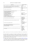
583 SUPPRESSION OF ITCHING BY THREE HERBAL ETHANOLIC EXTRACTS for A houstonianum, 203 nm for B falcatum, or 220 nm for S chinensis. The mobile phase was acetonitrile/methanol/water (30:40:30) for A houstonianum, 35% acetonitrile aqueous solution for B falcatum, and 65% acetonitrile aqueous solution for S chinensis. The column temperature was set at 35°C. CELL CULTURE The human keratinocyte cell line HaCaT was obtained from the Cell Lines Service (Eppelheim, Germany). HaCaT cells were maintained in Dulbecco’s modified Eagle medium supplemented with 10% fetal bovine serum (HyClone, Logan, UT, USA). CYTOTOXICITY ASSAY HaCaT cells were seeded into 96-well plates at a density of 5 × 10³ cells/well. After incubating for 24 h, cells were treated with vehicle (dimethyl sulfoxide) or various concentrations (10, 20, 40, and 80 μg/mL) of A houstonianum, B falcatum, and S chinensis for 24 h or a combination of the three (40 μg/mL A houstonianum, 20 μg/mL, and 40 μg/ mL S chinensis) for various time periods (0–48 h). Cytotoxicity was analyzed using a water- soluble formazan colorimetric assay-based Cell Counting Kit-8 (CCK-8 Dojindo Molecular Technologies, Gaithersburg, MD, USA) according to the manufacturer’s instructions. The absorbance at 450 nm was measured on an EMax Endpoint enzyme-linked immunosorbent assay (ELISA) Microplate Reader (Molecular Devices, Sunnyvale, CA, USA). REVERSE TRANSCRIPTION-POLYMERASE CHAIN REACTION Isolation of total RNA and synthesis of cDNA was performed as previously described (33). The reverse transcription (RT-PCR) primers and thermal cycling conditions were as follows: FLG forward, 5′-CAA ATC CTG AAG AAT CCA GAT GAC-3′, FLG reverse, 5′-TGC TTG AGC CAA CTT GAA TAC C-3′ (5 min at 94°C, followed by 30 s at 94°C, 35 s at 62°C, and 1 min at 72°C for 30 cycles) IL31 forward, 5′-GTC TTG GTA TTT ATG GAA TGC-3′, IL31 reverse, 5′- CCA GGG AGC ATT GAC AAC TCT TAG-3′ (5 min at 94°C, followed by 30 s at 94°C, 35 s at 55°C, and 1 min at 72°C for 30 cycles) POMC forward, 5′-CCT GCC TGG AAG ATG CCG AGA T-3′, POMC reverse, 5′-TGC TGC CGC TGC TGC TGC TGT-3′ (5 min at 94°C, followed by 30 s at 94°C, 35 s at 62°C, and 1 min at 72°C for 30 cycles) GAPDH forward, 5′-CCA AGG AGT AAG AAA CCC TGG AC-3′, GAPDH reverse, 5′-GGG CCG AGT TGG GAT AGG G-3′ (5 min at 94°C, followed by 30 s at 94°C, 30 s at 58°C, and 1 min at 72°C for 30 cycles) TSLP forward, 5’-TAG CAA TCG GCC ACA TTG CCT-3’, TSLP reverse, 5’-GAA GCG ACG CCA CAA TCC TTG -3′ (5 min at 94 followed by 30 s at 94°C, 35 s at 58°C, and 1 min at 72°C for 30 cycles). The amplified products were electrophoresed on a 2% agarose gel and visualized upon staining with ethidium bromide under ultraviolet light. QUANTITATIVE REAL-TIME PCR Validated commercial quantitative real-time PCR (qPCR) primers and SYBR Green-based fluorescent probes specific for FLG (id: qHsaCEP0039328), IL31 (id: qHsaCEP0055722),

584 JOURNAL OF COSMETIC SCIENCE TSLP (id: qHsaCIP0030468), and GAPDH (id: qHsaCEP0041396) were obtained from Bio-Rad (Hercules, CA, USA). The cycling conditions were as follows: denaturation at 95°C for 2 min, followed by 40 cycles using a step program (95°C for 10 s and 60°C for 45 s), as described previously (33). The relative expression levels of FLG, IL31, TSLP, and POMC mRNA were normalized to GAPDH mRNA levels using the software program provided by the manufacturer. IMMUNOBLOT ANALYSIS Whole-cell lysates were prepared by lysing HaCaT cells in a buffer containing 50 mM Tris-HCl (pH 7.4), 1% NP-40, 0.25% sodium deoxycholate, 500 mM NaCl, 1 mM ethylenediaminetetraacetic acid, 1 mM Na 3 VO 4 , 1 mM NaF, 10 μg/ml leupeptin, and 1 mM phenylmethylsulfonyl fluoride. Equal amounts of lysates were separated by sodium dodecyl sulfate-polyacrylamide gel electrophoresis and transferred onto 0.45 μm nitrocellulose membranes (Amersham Bioscience, Pittsburgh, PA, USA), as described previously (33). After blocking with 5% skimmed milk, the membrane was incubated with appropriate primary and secondary antibodies, and immunoreactive bands were detected using an enhanced chemiluminescence detection system (GE Healthcare, Piscataway, NJ, USA). The intensities of the immunoreactive bands were measured using ImageJ (version 1.52a National Institutes of Health, Bethesda, MD, USA). Target protein levels were normalized and plotted as fold-changes relative to the untreated control value. ELISA HaCaT cells were serum-starved for 24 h and then treated with 20 ng/mL IL4. The culture medium was collected after 24 h of incubation and the concentrations of IL31 and β-endorphin were determined using commercially available human IL31 ELISA (BioLegend, Cat #445704) and human β-endorphin ELISA kits (Biovision, Cat #E4458) following the manufacturer’s instructions. Briefly After attachment of the capture antibody to a 96-well plate, standards and samples were incubated with the capture antibody overnight at 4°C. Unbound samples were removed by washing detection antibodies that bind to each sample protein were added and the plate was incubated for 1–2 h at 25°C. Then, avidin-conjugated horseradish peroxidase solution was added, and the plate was again incubated for 30 min at 25°C, followed by the addition of substrate solution and incubation for 15 min in the dark. The absorbance at 450 nm was measured on an EMax Endpoint ELISA Microplate Reader (Molecular Devices). DOUBLE IMMUNOFLUORESCENCE STAINING HaCaT cells cultured on coverslips were left untreated or treated with IL4 (for IL31 expression) or IL4 plus IL13 (IL4 + IL13) (for FLG expression). After 24 h, the cells were fixed in 4% paraformaldehyde, permeabilized in 0.1% Triton X-100 and 2% bovine serum albumin, and incubated with primary antibodies against FLG and IL31. For FLG staining, the cells were incubated with anti-FLG (1:100) antibodies for 2 h, followed by Alexa Fluor 488-conjugated secondary antibodies (green fluorescence) for 30 min. F-actin was counterstained with rhodamine-labeled phalloidin (1:200 red fluorescence).
Purchased for the exclusive use of nofirst nolast (unknown)
From: SCC Media Library & Resource Center (library.scconline.org)