585 SUPPRESSION OF ITCHING BY THREE HERBAL ETHANOLIC EXTRACTS For IL31 staining, the cells were incubated with anti-IL31 (1:100) and antitubulin (1:200, for counterstaining) antibodies for 2 h, followed by Alexa Fluor 555-conjugated (red fluorescence for anti-IL31 antibody) and Alexa Fluor 488-conjugated (green fluorescence for antitubulin antibody) secondary antibodies for 30 min. For staining nuclear DNA, the cells were incubated with Hoechst 333258 (blue fluorescence) for 10 min. Fluorescent images were captured using an EVOS fluorescence microscope (Advance Microscopy Group, Bothell, WA, USA). CLINICAL EVALUATION PREPARATION OF TOPICAL CREAM FOR CLINICAL TRIAL The A houstonianum, B falcatum, and S chinensis mixture cream for the clinical test was prepared by Hansol Bio Company (Seongnam, Republic of Korea) in a Cosmetic Good Manufacturing Practice facility. The cream was prepared by mixing 2% A houstonianum, B falcatum, and S chinensis with a basal cream containing distilled water, butylene glycol, hydrogenated polyisobutene, ethylhexyl palmitate, octyldodecanol, glycerin, cetyl alcohol, sodium polyacryloyldimethyl taurate, polysorbate 60, dipropylene glycol, xylityl glucoside, xylitol, anhydroxylitol, beta-glucan, glyceryl stearate, hydrogenated polydecene, sorbitan stearate, glucose, allantoin, Trideceth-10, dipotassium glycyrrhizate, PEG-100 stearate, arginine, disodium EDTA, hydroxyacetophenone, caprylyl glycol, 1,2-hexanediol, ethylhexylglycerin, eucalyptus leaf oil, and limonene. PARTICIPANTS AND STUDY PROTOCOL A total of 33 female volunteers ages 20–65 with itchy skin and impaired skin barrier function (transepidermal water loss (TEWL) 12 g/m2/h) were included. Dermatologists evaluated and recorded the degree of erythema, scaling, induration, and fissuring, scoring 0 to 3 points at each participant’s visit, with a total of four visits per participant. This resulted in a possible total of 0 to 12 points if a participant’s total exceeded 6 points, they were judged as a subject requiring medical treatment and excluded from the study. The participants did not receive antibiotics, steroids, immunosuppressants, antihistamines, retinoids, or phototherapy for at least for 1 week prior to the clinical study. The study procedures were clearly explained to all selected subjects before they signed the informed consent form. The test creams were randomly marked “left” or “right,” so control site (untreated site) and tested site were randomized. Participants were instructed to apply 1 g or more of the provided test cream to the area 3 cm below the center of the inner elbow twice a day in the morning and evening. Participants visited the institution on days 0, 14, and 28 after the trial to measure TEWL, moisture content, and itching intensity. Subjects were instructed not to use body care products, showering, or bathing 12 h prior to the test. At the institution visited, participants were first placed at rest for 30 min in a room with controlled temperature (22±2°C) and humidity (50±10%). In addition, the investigator obtained written consent from each subject not to use the test cream on the control area to prevent unauthorized use. Volunteer participants were recruited by the Korea Dermatology Research Institute (Seongnam, Republic of Korea). The study was conducted under the Korean Cosmetics Good Clinical Practice (GCP) guidelines (approved IRB number: KDRI-IRB-21540).

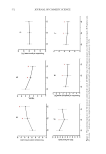
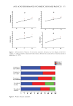
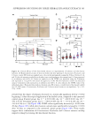
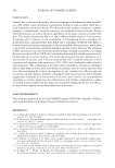
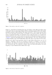
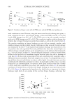
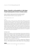
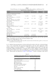
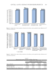
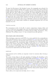
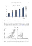
586 JOURNAL OF COSMETIC SCIENCE MEASUREMENTS TEWL was measured using a Tewameter TM-300 (Courage+Khazaka electronic GmbH, Germany). The results were expressed in g/m2/h. Skin moisture was assessed by measuring an electrical capacitance of the skin using a Corneometer CM825 (Courage+Khazaka electronic GmbH, Germany) in the laboratory. Moisture content was displayed digitally in arbitrary units (A.U.). Subjects self-recorded the degree of itchiness on the visual analog scale, ranging from 0 to 10 (No itching: 0 Mild itching: 1–less than 3 Moderate frequent itching: 3–less than 7 Severe itching: 7–less than 9 Very severe itching: 9–10) over the past 24 h at each institution visit on days 0, 14, and 28 days. STATISTICAL ANALYSIS Data are presented as the mean ± standard deviation (n = 3 for qPCR and ELISA data) or mean ± standard error of the mean (n = 33 for clinical trial data). Statistical analysis was performed using one-way analysis of variance followed by Dunnett’s or Sidak’s multiple comparisons test using GraphPad Prism (version 9.2.0 GraphPad Software, San Diego, CA, USA). For all analyses, differences with p 0.05 were considered statistically significant. RESULTS AND DISCUSSION ANALYTICAL HPLC PROFILES We performed HPLC analysis using an Agilent 1260 Infinity system to characterize the phytochemical components of the ethanolic extracts. We used precocene II, saikosaponin A, and gomisin A/gomisin N/angeloylgomisin H as reference compounds in A houstonianum, B falcatum, and S chinensis, respectively. The chromatograms of A houstonianum, B falcatum, and S chinensis were compared by the retention time to that of the reference standard compound (Figure 1). These data suggest that A houstonianum, B falcatum, and S chinensis contained active phytochemical compounds. ETHANOLIC EXTRACTS OF THE THREE DIFFERENT HERBS DO NOT INDUCE HACAT CYTOTOXICITY First, we determined the cytotoxicity of the three herbal ethanolic extracts. HaCaT cells were treated with different concentrations (0–80 μg/mL) for 24 h, and a cell viability assay was performed. Treatment with A houstonianum, B falcatum, or S chinensis did not induce considerable cytotoxicity up to 80 μg/mL compared to the vehicle-treated control (0 μg/mL) (Figure 2A). A combination of the three extracts showed no significant cytotoxicity until at least 48 h after treatment (Figure 2B). In the following experiments, we routinely used the combination of 40 μg/mL A houstonianum, 20 μg/mL B falcatum, and 40 μg/mL S chinensis. THREE HERBAL EXTRACTS RECOVER FLG EXPRESSION REDUCED BY IL4 + IL13 STIMULATION AD is one of the most common chronic inflammatory skin diseases characterized by prolonged and intense itching (34), making it ideal for studying the pathophysiology of pruritus. The well-known genetic risk factor for AD is a null mutation in the gene encoding
Purchased for the exclusive use of nofirst nolast (unknown)
From: SCC Media Library & Resource Center (library.scconline.org)