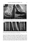
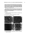
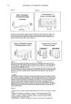
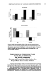
j. Cosmet. sci., 49, 13-22 (January/February 1998) Analysis of the damaged components of permed hair using biochemical technique R. KON, A. NAKAMURA, N. HIRABAYASHI, and K. TAKEUCHI, Analytical Research Center, Lion Corp., Hirai 7-13-12, Edogawa-ku, Tokyo 132, Japan. Accepted for publication September 30, •997. Presented at the 38th Scientific Meeting of the Society of Cosmetic Chemists of Japan, Osaka, June 18, 1996. Synopsis We have developed a new method of fractionating hair components in order to analyze the damaged components and the degree of damage due to perming. We found that the amount of constituent proteins extracted by an anionic surfactant with reductant was influenced by the concentration of the reductant. Using this method, the matrix and the microfibril protein could be easily separated and quantified. Applying this method to the analysis of individual hairs, we found a significant decrease in the "intact" microfibril protein on the tip end of permed hair. INTRODUCTION Many cosmetic investigators are studying ways to evaluate the degree of hair damage or to develop anti-damaging products, but hair is a complex organization and the damage can first be seen at the hair's tip, which may have been on the head 2-3 years and damaged by cumulative cosmetic behavior. Therefore, efforts to confirm the correlation between damage and cause are very difficult. It has been reported that hair is damaged by various causes such as sunlight (1), grooming (shampooing, drying, brushing, and combing) (2), and cosmetic treatments (perming, dyeing, and bleaching) (3). Robbins concluded that the cause of hair damage is the sum of all grooming practices (rubbing, stretching, and washing), while sunlight or chemical processing treatments make the hair more susceptible to such damaging actions (4). However, these studies mainly dealt with mechanical properties or morphological changes such as decreasing cuticle layers, scale lift, and split ends, examined by electron microscopy. A few investigators have studied the alteration of the total amino acids or lipid compositions (5,6). The proteins of hair constitute 80% or more of the total mass, but studying these proteins is difficult because of their insolubility due to the disulfide-bonded polymeric structure. Consequently, a few investigations were reported (7,8), but it is still unknown 13

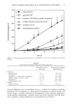
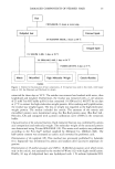
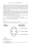
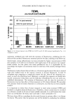
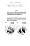
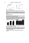
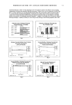
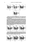
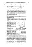
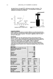
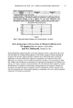
14 JOURNAL OF COSMETIC SCIENCE how the hair components, especially proteinous molecules, change due to perming. Generally, the hair proteins are extracted by reductants and denaturing agents such as urea or guanidine. Further isolation of the main classes of hair proteins (the matrix and the microfibril) requires an isoelectric precipitation step or electrophoretic method (9). This method is not of practical use for the analysis of individual hairs because it requires many hairs and is a complicated method. Thus, we attempted to develop a simple and easy extraction method for quantifying hair components using a reductant with an anionic surfactant. The goal of this study is the analysis of damage to an individual hair due to perming. MATERIALS AND METHODS CHEMICALS Analytical grade 2-mercaptoethanol (2-ME), dithiothreitol (DTT), tricine, and SDS were supplied by Nacalai Tesque, Inc. (Kyoto, Japan). 4-Bromomethyl-7-methoxycoumarin was purchased from Dojindo Laboratories, Inc. (Kumamoto, Japan). •/-Glutamyl-g- lysine (isopeptide), pronase, leu-aminopeptidase, prolidase, and carboxypeptidases were purchased from the Sigma Chemical Co. (St. Louis, MO). HAIRS For the development of the methods, we collected the root end hair from ten Japanese women who had not been exposed to chemical treatments such as perming or dyeing. Hair was washed using 1% (w/w) SDS and then thoroughly rinsed with tap water. The hair was dried, and external lipids were extracted for 16 hours at room temperature in a 100-fold solution of chloroform/methanol (2:1 v/v). The delipidized hair was cut into 10-mm sections. For the analysis of an individual hair, we collected 100 fibers ofpermed hair from two Japanese women who had permed every two or three months. One hundred hairs that had not been exposed to cosmetic treatments such as perming or dyeing were also collected from one Japanese woman. The hair was cut into 100-mm lengths from the root end to the tip end and then treated as described. METHODS Extraction of hair proteins. The hair proteins were extracted using an anionic surfactant with reductant. In detail, a 10-mg sample of delipidized hair was immersed in 1 ml of 25 mM Tris-HC1 buffer (pH 8.3) that contained 1% SDS and various concentrations of 2-ME. The protein was extracted for three days at 50øC. The extracted protein was analyzed using Tricine-SDS-PAGE (10). Fractionation of hair components. The hair components were fractionated according to the scheme illustrated in Figure 1. A 10-mg sample of delipidized hair was immersed in a 1-ml solution of 25 mM Tris-HC1 buffer (pH 8.3) that contained 1% SDS and 2 M 2-ME. The matrix protein was extracted for three days at 50øC. The hair was washed with 25 mM Tris-HCl buffer, then immersed in a 1-ml solution of 25 mM Tris-HC1 buffer (pH 8.3) that contained 1% SDS and 0.4 M 2-ME. The microfibril protein was
Purchased for the exclusive use of nofirst nolast (unknown)
From: SCC Media Library & Resource Center (library.scconline.org)