PERCUTANEOUS DIFFUSION OF A HYDROPHILIC SUNSCREEN 11 (8) M.J. Cappel and J. Kreuter, Effect of nonionic surfactants on transdermal drug delivery: I. Polysor- bates, In. J. Pharm., 69, 143-153 (1991). (9) K.A. Walters, M. Walker, and O. Olejnik, Non-ionic surfactant effects in hairless mouse skin permeability characteristics, J. Pharm. Pharmaco/., 40, 525-529 (1988). (10) Z.T. Chowhah and R. Pritchard, Effect of surfactants on percutaneous absorption of naproxen. I. Comparisons of rabbit, rat, and human excised skin,.]. Phar•. Sci., 67, 1272-1274 (1978). (11) W. W. Shen, A. G. Danti, and F. N. Bruscato, Effect of nonionic surfactants on percutaneous absorp- tion of salicylic acid and sodium salicylate in the presence of dimethyl sulfoxide, J. Phar•. Sd., 65, 1780-1783 (1976). (12) H. G. Ibrahim, Release study from lyotropic liquid crystal systems,J. Pharm. Sd., 78,683-687 (1989). (13) H. L. G. M. Tiemessen, H. E. Bodde, C. VanMourik, and H. E. Junginger, In vitro drug release from liquid crystalline creams: Cream structure dependence, Progr. Colloid. Polymer Sci., 77, 131-135 (1988). (14) C. C. Muller-Goymann and S. G. Frank, Interaction of lidocaine-HC1 with the liquid crystal structure of topical preparations, Int..]. Pharm., 29, 147-159 (1986). (15) S. Wahlgren, A. L. Linsdtrom, and S. E. Friberg, Liquid crystals as a potential ointment vehicle,.]. Pharm. Sci., 73, 1484-1486 (1984). (16) K. Kriwet and C. C. Muller-Goymann, Diclofenac release from phospholipid drug systems and per- meation through excised human stratum corneum, Int..]. Pha•m., 125, 231-242 (1995). (17) I.L. Wilisch and C. C. Muller-Goymann, Correlation of colloidal microstructure, drug release and permeation through excised human skin, Int..]. Pharm., 96, 79-84 (1993). (18) J. Swarbrick and J. Siverly, The influence of liquid crystalline phases on percutaneous drugs. II. Percutaneous studies through excised human skin, Pharm. Res., 9, 1550-1555 (1992). (19) D. W. Osborne, A.J.I. Ward, and K.J. O'Neill, "Surfactant Association Colloids as Topical Drug Delivery Vehicles," in Topical Drug Delivery Formulations, D. W. Osborne and A. H. Amann, Eds. (Marcel Dekker, New York, 1990), pp. 349-379. (20) S. B. Ruddy, "Surfactants," in Percutaneous Penetration Enhancers, E. W. Smith and H. I. Maibach, Eds. (CRC Press, Boca Raton, FL, 1995), pp. 245-257. (21) P. Ashton, J. Hadgraft, and K. A. Walters, Effects of surfactants in percutaneous absorption, Pharm. Acta Helv., 61, 228-235 (1986). (22) K.A. Walters, "Surfactants and Percutaneous Absorption," in Prediction of Percutaneous Penetration, R. C. Scott, R. H. Guy and J. Hadgraft, Eds. (IBC Technical Services, London, 1989) pp. 148-162. (23) D. Atwood, A. T. Florence, Surfactants Systems (Chapman and Hall, London, 1983) pp. 124-228. (24) P. P. Sarpotdar and J. L. Zatz, Evaluation of penetration enhancement of lidocaine by nonionic sur- factants through hairless mouse skin in vitro, .]. Pharm. Sci., 75, 176-181 (1986). (25) M. Loden, The simultaneous penetration of water and sodium lauryl sulfate through isolated human skin,.]. Soc. Cosmet. Chem., 41, 227-233 (1990). (26) J. L. Zatz, "Modification of Skin Permeation by Surface-Active Agents," in Skin Permeation: Funda- mentals and Application, J. L. Zatz, Ed. (Allured Publishing Corp., Wheaton, IL, 1993), pp. 149-162. (27) P.M. Elias, Epidermal lipids, membranes, and keratinization, Int..]. Dermatol., 20, 1-19 (1981). (28) B. Ongpipattanakul, M. L. Francoeur, and R. O. Potts, Polymorphism in stratum corneum, Blochim. Biophys. Acta, 1190, 122-155 (1994). (29) D.T. Parrott and J. E. Turner, Mesophase formation by ceramides and cholesterol: A model for stratum corneum lipid packing?, Blochim. Biophys. Acta, 1147, 273-276 (1993). (30) S.J. Rehfeld, W. Z. Plachy, S. Y. E. Hou, and P.M. Elias, Localization of lipid microdomains and thermal phenomena in murine stratum corneum and isolated membrane complexes: An electron spin resonance study,.]. Invest. Dermatol., 95,217-223 (1990). (31) E.J. French, C. W. Pouton, and G. Steele, "Fluidization of Lipid Bilayers by Non-Ionic Surfactants: Structure-Activity Studies Using a Fluorescent Probe," in Prediction of Percutaneous Penetration, R. C. Scott, R. H. Guy, J. Hadgraft, Eds. (IBC Technical Services, London, 1989), pp. 308-315. (32) H. E.J. Hofland, J. A. Bouwstra, F. Spies, H. E. Bodde, J. F. Nagelkerke, C. Cultander, and H. E. Junginger, Interactions between non-ionic surfactant vesicles and human stratum corneum in vitro,.]. Liposome Res., 5, 241-263 (1995). (33) J. Lasch and J. Bouwstra, Interactions of external lipids (lipid vesicles) with the skin,.]. Liposome Res., 5, 543-569 (1995).

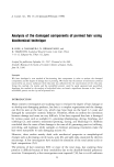
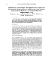
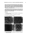
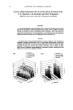
j. Cosmet. sci., 49, 13-22 (January/February 1998) Analysis of the damaged components of permed hair using biochemical technique R. KON, A. NAKAMURA, N. HIRABAYASHI, and K. TAKEUCHI, Analytical Research Center, Lion Corp., Hirai 7-13-12, Edogawa-ku, Tokyo 132, Japan. Accepted for publication September 30, •997. Presented at the 38th Scientific Meeting of the Society of Cosmetic Chemists of Japan, Osaka, June 18, 1996. Synopsis We have developed a new method of fractionating hair components in order to analyze the damaged components and the degree of damage due to perming. We found that the amount of constituent proteins extracted by an anionic surfactant with reductant was influenced by the concentration of the reductant. Using this method, the matrix and the microfibril protein could be easily separated and quantified. Applying this method to the analysis of individual hairs, we found a significant decrease in the "intact" microfibril protein on the tip end of permed hair. INTRODUCTION Many cosmetic investigators are studying ways to evaluate the degree of hair damage or to develop anti-damaging products, but hair is a complex organization and the damage can first be seen at the hair's tip, which may have been on the head 2-3 years and damaged by cumulative cosmetic behavior. Therefore, efforts to confirm the correlation between damage and cause are very difficult. It has been reported that hair is damaged by various causes such as sunlight (1), grooming (shampooing, drying, brushing, and combing) (2), and cosmetic treatments (perming, dyeing, and bleaching) (3). Robbins concluded that the cause of hair damage is the sum of all grooming practices (rubbing, stretching, and washing), while sunlight or chemical processing treatments make the hair more susceptible to such damaging actions (4). However, these studies mainly dealt with mechanical properties or morphological changes such as decreasing cuticle layers, scale lift, and split ends, examined by electron microscopy. A few investigators have studied the alteration of the total amino acids or lipid compositions (5,6). The proteins of hair constitute 80% or more of the total mass, but studying these proteins is difficult because of their insolubility due to the disulfide-bonded polymeric structure. Consequently, a few investigations were reported (7,8), but it is still unknown 13
Purchased for the exclusive use of nofirst nolast (unknown)
From: SCC Media Library & Resource Center (library.scconline.org)