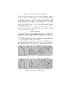
HYPO- AND HYPERPIGMENTATION OF SKIN :"•i'i' basal cells (4). Masson and his group believed the origin of the pigment- forming dendritic cells and cellules claires to be neural in origin, migrating from the neural crest to the epidermis during embryohal life (5). This .::..later theory has won many converts and appears to be most popularly ac- i:(• cepted today. :":' .•. The actual study of histochemical and enzymatic reactions in melanin production was given impetus by the historic Dopa Reaction (4). Sections of skin placed in Dopa [•-3,4-(dihydroxyphenyl)-L-alanine] for 24 hours at 37øC. and examined under the microscope showed a darkening in the basal cell layer. The cells that stained dark were called dopa-positive. 'Bloch believed that the pigment producing cells contained the specific enzyme dopa-oxidase which changed the colorless dopa into an insoluble :".colored product, dopa melanin. Further work by other authors has shown •.. that dopa also could be converted to melanin by other enzymes. Tyrosinase has been recently identified as the main enzyme in melanin production. Its presence in lower animal and plant life has been demon- strated(6) and its occurrence in human skin has been shown (7). Tyrosine is activated by tyrosinase to produce dopa which in turn is carried through a series of intermediates to finally produce melanin. This conversion reaction is markedly accelerated by small amounts of dopa. Tyrosinase Tyrosinase Tyrosine ) Dopa ) (Intermediates) ) Melanin Dopa Oxidase In experimental studies performed (8), it was shown that in normal non- irradiated human epidermis, tyrosinase exists in an inactive state and can- not convert tyrosine to melanin. Upon radiation, the system becomes active and melanin is formed. Why radiant energy activates the enzyme reaction isn't known but Fitzpatrick offered two possible explanations: (1) traces of dopa which greatly accelerate the tyrosin-tyrosinase reaction may be formed in the melanocyte by the ultraviolet energy and thus catalyze the reaction or (2) the concentration of the S--H groups normally occur- ring in the epidermis and which inhibit tyrosinase activity by combining with copper may be decreased. Biopsies of human skin previously exposed to ultraviolet light radiation in vivo were incubated in a tyrosine-phosphate buffer and melanocytes were found in the basal cell layer at the epidermal-dermal junction. In control sections of non-irradiated skin and irradiated skin which was not placed in the tyrosine substrate, no melanocyte activity was seen. The tyrosine-tyrosinase system was also shown to exist in the "inhibited" state in benign pigmented skin lesions such as nevi (moles) and ephilides (freckles). In malignant melanoma, however, as with normal irradiated melanocytes, an active system prevails. This lends itself to further in- vestigative study for a chemical means of differentiation of benign from malignant pigmented lesions (8).

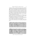
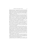
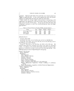
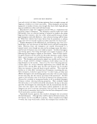
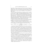
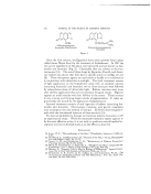
248 JOURNAL OF THE SOCIETY OF COSMETIC CHEMISTS INHIBITION OF MELANIN FORMATION Hyperpigmentation due to melanin has always been a fascinating but disheartening problem. Attempts to interfere in vivo with melanization or to decrease pigmentation have been far from consistently successful. The so-called bleaching or freckle creams have been used for years. Prep- arations containing such ingredients as resorcinol, salicylic acid, potas- sium hydroxide, and acetic acid are aimed at reducing pigmentation by exfoliation. Oxidizing agents such as hydrogen peroxide and perborates are used with minimal effectiveness. Mercury preparations of which am- moniated mercury creams are the most popular still have wide usage der- matologically. Nealon has found mercury to be quite effective and has shown its skin lightening effects in a series of cases (9). Although previous efficacy was believed due to its peeling action, evidence points toward its direct interference by means of competing with copper ions present in the skin for an active center on the enzyme tyrosinase to form an inactive en- zyme (10). The role of Vitamin C in pigmentation is probably also related to adrenal activity. The pigmentation seen in Addison's disease has been lessened in some cases by large oral doses of ascorbic acid (11). Vitamin C may also help to keep melanin in a reduced state which is light brown in color or by inhibiting the oxidation of dopa quinone. In 1936 Oettal fed hydroquinone to black-haired cats in a period of 6-8 weeks, their hair turned gray. After the drug was stopped, the hair re- gained its color (12). In 1939 occupational leukoderma due to an antioxidant in rubber gloves was reported (13). Since then other cases oldepigmentation of the skin in contact with the monobenzylether of hydroquinone have appeared in the literature. In almost all instances, the pigment has returned. OH OH OH , , OH O C--O Hydroquinone [ I CH• C•Hs i p-Hydroxy- (• propiophenone Monobenzylether of Hydroqui none Figure 1
Purchased for the exclusive use of nofirst nolast (unknown)
From: SCC Media Library & Resource Center (library.scconline.org)