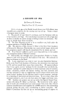
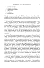
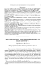
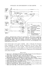
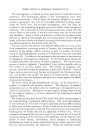
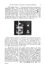
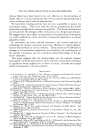
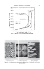
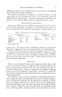
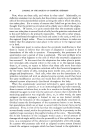
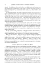
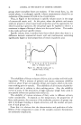
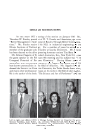
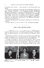
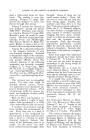
PHYSIOLOGY AND HISTOCHEMISTRY OF HAIR GROWTH 13 -- k, I '• comeurn • -- L '• sebum ,j mertt• lrtto new ' -rn , K I "-. dub Figure 1.--Postulated skin and hair cycle control diagram. BE, basal layer of epidermis 2PSC, peripheral cells of sebaceous gland UES, upper permanent external sheath D/•, derreal papilla CTS, connective tissue sheath C, corium z/, adipose layer /•H, pig- ment cells of hair follicle LES, lower temporary external sheath m, mitotic activity k, keratinization l, lipid accumulation g, glycogen accumulation mel, melanogenesis hrhr, hairless genotype of the mouse. more precise than can be done verbally. Also it indicates the reactions most susceptible to possible outside disturbances and the areas where further investigations are most needed. There are general, systemic fac- tors, such as hormones, or food supply, but also there are pronounced local conditions and reactions which determine to a large extent the maintenance and cycles of the skin. BIBLIOGRAPHY (1) Argyris, T. S., "The Relationship Between the Hair Growth Cycle and the Response of Mouse Skin to X-irradiation," •Im. •e. •Inat., 94, 439 (1954). (2) Chase, H. B., "Growth of the Hair," 2Physio/. Rev., 34, 112 (1954). (3) Chase, H. B., "Evaluation of Biological Control Systems," Tech. Report: "Essays on Biological Utilization," Control Systems Lab., University of Illinois (1954). (4) Chase, H. B., and Montagna, W., "Relation of Hair Proliferation to Damage Induced in the Mouse Skin," 2Proc. Soc. Expt/. Bio/. Med., 76, 35 (1951). (5) Chase, H. B., Montagna, W., and Malone, J. D., "Changes in the Skin in Relation to the Hair Growth Cycle," •Inat. Record, 116, 75 (1953). (6) Chase, H. B., Rauch, H., and Smith, V. W., "Critical Stages of Hair Development and Pigmentation in the Mouse," 2Physiol. Zool., 24, 1 (1951). (7) Eisen, A. Z., Montagna, W., and Chase, H. B., "Sulfhydryl Groups in the Skin of the Mouse and Guinea Pig," •7. Nat. Cancer Inst., 14, 341 (1953).

14 JOURNAL OF THE SOCIETY OF COSMETIC CHEMISTS (8) Liang, H., and Cowdry, E. V., "Changes of Hair Follicular Cells After a Single Painting of Methylcholanthrene in Mice," Cancer Research, 14, 340 (1954). (9) Loewenthal, L. A., "The Effects of Vitamin A Deficiency on Skin and Hair Growth in Mice." Thesis, Brown University (1954). (10) Montagna, W., and Chase, H. B.• "Redifferentiation of Sebaceous Glands in the Mouse After Total Extirpation with Methylcholanthrene," .4nat. Record, 107, 83 (1950). (11) Montagna, W., Chase, H. B., Malone, J. D., and M, elaragno, H. P., "Cyclic Changes in Polysaccharides of the Papilla of the Hair Follicle, ' Quart. •. Microgiol. $ci., 93, 241 (1952). (12) Pinkus, H., "Examination of the Epidermis by the Strip Method, II. Biometric Data on Regeneration of the Human Epidermis," f. Investigative DermatoL, 19, 431 (1952). NEWER ASPECTS OF EPIDERMAL D I F F ERENTIATI ON* By PETER FLY. Sell, M.D., PH.D. Department of Dermatology, University of Pennsylvania School of Medicine, Philadelphia, Pa. HUMAN EPIDERMIS, the outermost layer of the skin, is in most areas only about 1/i000 of an inch deep. The thickness of an onion skin paper is all that separates the inner from the outer environment. Within this layer an amazing variety of changes occur, as the epidermal cells differentiate into the dead horny layer at the surface. Some of these changes are environ- mental, such as a drop in pH from the inner alkaline tissue fluids to the acid reaction of the surface film the temperature and water content also de- crease as one proceeds from the inside to the outside. The most character- istic changes, however, are inherent in the epidermal cells themselves. These changes may be classified as morphological, physical, and chemical. Such a classification is arbitrary terms such as "morphological, .... physi- cal," or "chemical" denote different aspects of the same process, or are dif- ferent ways of looking at the same thing. The time is not far away, indeed in many instances has already arrived, when it will become possible to ex- press morphological features in chemical or physical terms. Among the morphological changes the conversion of the epidermal cells into the fibrous structures of the horny layer is the most conspicuous one. This transformation is a continuous and slow process it has been esti- mated that under normal circumstances at least a month, or probably more time, elapses before a cell in the basal layer reaches full maturity and is cast off as a horny scale of microscopic dimensions on the surface of the skin (1). The morphological details of this conversion are not very clear. * Presented at the December 9, 1954, Meeting, New York City. This study was supported by U.S. Public Health Grant #G4257.
Purchased for the exclusive use of nofirst nolast (unknown)
From: SCC Media Library & Resource Center (library.scconline.org)