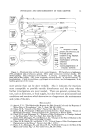
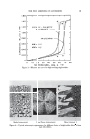
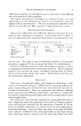
NEWER ASPECTS OF EPIDERMAL DIFFERENTIATION 15 Keratinization, like collagen formation (2), probably begins in the periph- ery of the cell and proceeds from there throughout the rest of the cyto- plasm (1). Studies of a so-called transitional layer, the granular layer, which is located immediately underneath the horny layer, t did not shed much light on the morphological aspects of epidermal keratinization. The granular layer is composed of cells filled with a granular substance, woefully misnamed keratohyalin. Keratohyalin is neither keratin nor hyalin it is deplorable that the name implies that this material is a forerunner of keratin. Such an assumption is not borne out by the fact that some types of keratinization, such as nail formation, are characterized by the absence of a granular layer (3). In a pathologic type of keratinization, called para- keratosis, nuclear remnants are retained in the horny layer and the granular layer is absent. Although by no means proved, it would appear that an accelerated rate of keratinization leads to failure of keratohyalin granules to appear. The most likely explanation of the origin of the keratohyalin granules is that they are by-products in the keratinization process. As the cytoplasm partly solidifies into keratin and partly disintegrates, some of the intracellular proteins may be precipitated and give the appearance of granules (4). The disintegration of the cytoplasm and unmasking of the protein molecule immediately underneath the horny layer are also indicated by the sudden release of acid phosphatase (5) and by the frequent appear- ance of an increased sulfhydryl reaction in this region, also called the keratogenous zone (6). It has been postulated for a long time that from a physical standpoint the epidermis contains globular and fibrous proteins. The globular proteins are presumably involved in cellular metabolism, while the fibrous struc- tures represent the forerunners of the keratin fibers. A rather well-defined fibrous protein, epidermin, was extracted with 6 M urea from the super- ficial epidermal layers of the cow's nose. This protein had an x-ray diffrac- tion pattern like alpha keratin (7). In human epidermis, which has a struc- ture vastly different from the epidermis on the cow's nose (8), epidermin has never been shown to occur. Moreover, the process of extraction with 6 M urea, as used for epidermin, leads to denaturation-of epidermal pro- teins. Thus, the two distinct globular proteins extracted from human epidermis with neutral buffers become indistinguishable after treatment with 6 M urea (9). The characterization of epidermal proteins remains a task for the future, and is dependent on the availability of suitable solvents which can extract the more resistant fibrous structures in the epidermal cells without causing undue denaturation (8). From a chemical point of view, during epidermal differentiation, two proc- esses go on simultaneously: 'the formation or consolidation of the keratin 1 t The so-called stratum lucidurn, a layer between the granular and horny layers, can be visualized on the palms and soles only.

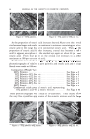
16 JOURNAL OF THE SOCIETY OF COSMETIC CHEMISTS fibers and the disintegration of structures which apparently do not directly contribute to the formation of keratin, such as the nucleus and some parts of cytoplasm. Keratinization of the epidermal cells results in the formation of a so- called soft keratin, which flakes off and is characterized by a relatively low cystine content. Very little is known about the chemical changes that occur during the synthesis of epidermal keratin. The scant analytical data on the amino acid composition of human epidermis and horny layer are listed in Table 1. TABLE I--AMINO ACID COMPOSITION OF HUMAN EPIDERMIS AND HORNY LAYE• Epidermis Horny layer Arginine Lysine Histidine Cystine Tyrosine•yptophane 6.7 5.9 2.6 1.4 4.0 1.0 5.9-10.0 3.1-7.1 0.6-1.9 2.3-3.8 3.•5.7 0.5-1.8 These data, originating from different authors (1, 10), are open to some criticism. "Epidermis" in the table stands for whole epidermis which comprises both cellular and keratinous components, while the horny layer analyzed represents material from a variety of normal and pathological sources. Moreover, methodological and biological variations may account for the variability of the results. With these reservations in mind, it would appear that during keratinization there is little change in the over-all amino acid composition, except for a possible reduction in the histidine con- tent and an increase in the concentration of cystine. In our own analyses of the horny and cellular layers from the same samples of human epidermis, the cystine values remained essentially the same, or showed a slight increase only, throughout the entire thickness of the epidermis (6, 11). This finding is of some theoretical interest. In the past, on the basis of inadequate histochemical evidence, it has been claimed that the oxidation of sulfhydryl groups to disulfide linkages played a major role in epidermal keratinization (12). This theory could not be confirmed either by recent histochemical studies (13) or by our direct chemi- cal analyses (6, 11, 14). The constancy of the cystine values throughout the entire thickness of the epidermis suggests that the consolidaton of the keratin fibers through disulfide bridge formation starts already in the deep cellular layers of the epidermis, and that there is no extensive disulfide bridge formation between the cellular and horny layers, as has been pre- viously thought. Of interest is also the recent finding that sulfhydryl groups (cysteine) are retained in all keratins studied, especially in epidermal keratins (6, 11). With an increasing rate of keratin formation, the concentration of these groups rises. The persistence of the sulfhydryl groups in the horny layer probably renders keratin more susceptible to chemical and physical in- fluences.
Purchased for the exclusive use of nofirst nolast (unknown)
From: SCC Media Library & Resource Center (library.scconline.org)