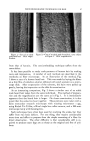
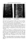
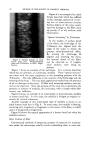
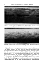
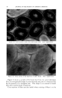
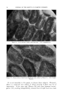
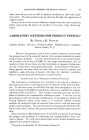
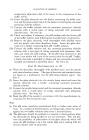
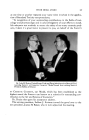
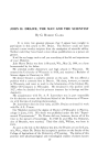
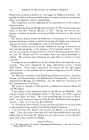
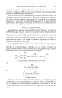
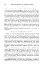
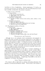
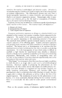
92 JOURNAL OF THE SOCIETY OF COSMETIC CHEMISTS Figure 9.--"Optical section" showing discontinuous medulla in human head hair. Photo- micrograph taken with Trilineator at 500X magnification. Figure 10.--"Optlcal section" of human head hair without medulla. Photomicrograph taken with Trilineator at 500X magnification. Figure 12 shows an example of a cross section prepared in this manner. The sample consists of ten hairs from the head of a dark-haired child. Since it was desired to observe specifically these ten hairs, they were packed into the microtome surrounded by bleached hair which could be distin- guished easily. This and the following three pictures were taken with the same equipment as the preceding photomicrographs, except that the Trilineator was not used. The film was Kodak Panatomic X. As a contrast, Fig. 13 shows some red hair from a female adult human head. The individual pigment granules are easier to see in this picture. To distinguish easily the ten fibers of the sample, they were packed in

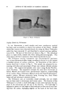
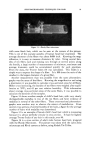
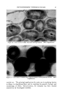
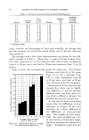
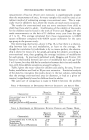
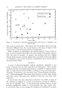
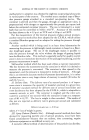
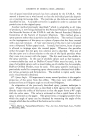
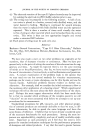
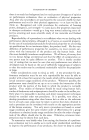
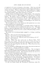
PHOTOMICROGRAPHIC TECHNIQUES FOR HAIR 93 Figure 11.--Hardy fiber microtome. with some black hair, which can be seen at the corners of the picture. This is one of the coarsest samples of human head hair examined. The average diameter of the fibers was about 0.004 inch. Knowing the mag- nification, it is easy to measure diameters by ruler. Using several bun- dles of ten fibers each and making cuts through at several points along the bundle, a large number of measurements of minimum, maximum and average diameters could be accumulated quickly for each specimen. From the data, the Trotter Index (9) was calculated. This Index is a simple way to express the shape of a fiber. It is 100 times the ratio of the smallest to the largest diameter of a given fiber. Another measurement that was possible from the same photomicro- graphs was the areas of the fibers. Knowing the magnification and using a planimeter, it was possible to calculate cross-sectional areas for all fibers. Before cross sectioning, the bundles of ten fibers were weighed after equili- bration at 70øF., and 65 per cent relative humidity. With information about average cross-sectional areas of the same fibers, it was possible to calculate the densities of the samples. Figure 14 shows another sample of child's head hair, with very clearly distinguishable medullas in two of the ten fibers and faint evidence of medullas in several of the other fibers. These cross-sectional photomicro- graphs were another way to observe the extent of medullation. From several cross sections of several bundles of ten hairs, a figure for the average number of hairs with medullas was determined. Figure 15 is an example of female child's head hair which is interesting because it is almost perfectly circular in cross section. It had the highest average Trotter Index of any hair in this study, over 90. Figure 16 is a cross section of adult male human head hair, prepared with the Hardy Microtome. The picture was taken with the same Zeiss microscope and Rival camera as before, using Kodak Tri-X film.
Purchased for the exclusive use of nofirst nolast (unknown)
From: SCC Media Library & Resource Center (library.scconline.org)