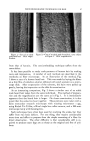
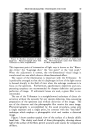
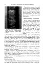
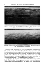
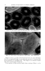
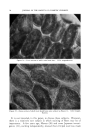
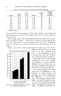
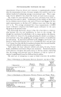
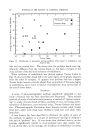
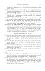
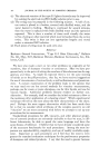
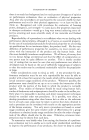
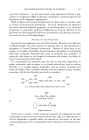
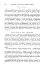
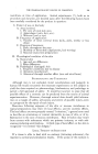
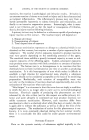
90 JOURNAL OF THE SOCIETY OF COSMETIC CHEMISTS Figure &--Surface damage on adult female head hair. Photomicrograph taken with Trilineator, at 500X magni- fication. Figure 6 is an example of an adult female head hair which has suffered surface damage, apparently involv- ing loss of some sections of scales. Surface defects of this type, result- ing from damage to the fiber, show up poorly, if at all, without relief illumination. "Optical Sectioning" by Trilineator In the studies of surface detail of the fibers, the knife-edge of the Trilineator was aligned with the edges of the scales to obtain the greatest three-dimensional effect. By turning the knife-edge 90 degrees and lowering the focus, the internal detail of the fibers can be observed as if longitu- dinal sections were being ex- amined. Figure 7 shows an example of this technique. It is a human head hair which has an unbroken, or continuous, medulla. These "optical sections" are taken with the same equipment as the preceding pictures with the Trilineator. The only difference is the rotation of the knife-edge and the lowering of the focus. The very dark appearance results from the presence of air or other gas in the canal. If filled with mounting medium, more de- tail can be observed. Since the purpose of this work was to determine the presence Or absence of medulla, this technique, with a simple whole fiber mount, was sufficient. Figure 8 shows an example of an interrupted, or discontinuous, medulla in human head hair. In this type, the medulla is bridged at irregular in- tervals by cortical substance. Another example of this interrupted type of medulla is shown in an adult human head hair in Fig. 9. In some cases, the medulla is fleeting, occurring only irregularly as fragments in the center of the fiber, in which case it is designated as fragmental. Figure 10 shows the typical appearance of a human head hair when the medulla is absent. Cross Sections of Hair Convkntional methods of Ereparing samples"0f materials for examina- tibn dhder the m•cx:oscope usually involve embedding them in some rrfk-

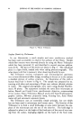
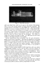
PHOTOMICROGRAPHIC TECHNIQUES FOR HAIR 91 Figure 7.--"Optical section" showing continuous medulla in human head hair. Photomicro- graph taken with Trilineator at 500X magnification. .. "•.:-•? ?•--.,... '-:..::.,, . - ............. .,::.,- ß ..... •---•. .-.• ....... ' ........ •"s-- ..x.....?- :• .... ---..-'.•,--...-.:F,':.?/?5-. ..... .x•:• '. : :•:•* •' - ..... .% -,' •- :*:: :,,v': '"'*' :'"." ...... . .... •- .- r-. "r '** """ : • '*'. .... Figure 8.•-"Optical section" showing discontinuous medulla in human he ad hair. Photo- •- micrograph taken with Trilineator at $00X magnification. terial, such as paraffin or some resin or polymer, and then slicing sections with a rotary microtome. These methods are not suitable for human hair because the fibers slip too easily out of the embedding material when the blade hits them. Accordingly, a variety of special microtomes have been devised for fibrous materials, and a description of them can be found in any of the textbooks on fiber microscopy. The simplest of these, and one which is quite suitable for work with human hair, is the Hardy Fiber Mi- crotome, which is shown in Fig. 11. To use this device, the two halves are disengaged, and a bundle of fiberõ' is inserted in the slot and held in pl&ce by inserting the tongue of the other half. The sample is'cut off flush with both faces of the plate, and the desired thickness of section is pushed out through the slot by means of a graduated micrometer screw •rrangement. Before slicing off the section" with a razor blade, the speci- men is cemented together with collodion. "'
Purchased for the exclusive use of nofirst nolast (unknown)
From: SCC Media Library & Resource Center (library.scconline.org)