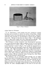
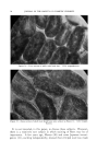
METHODS OF INCREASING SKIN PIGMENTATION 81 (daily sunlight, Addison's disease). In these conditions the melanocytes are only partially filled and the horny layer contains less than the maximum amount of melanin. It is possible that the, e is not enough substrate (tyrosine) available to maintain maximum pigment forming activity. This brief summary demonstrates that pigment formation must proceed for some time before a large amount of melanin is contained in the skin and that regardless of stimulation there is a limit to the amount of melanin that can be produced. Sunlight is the most frequently utilized pigmenting agent. The exact mechanism of action is unknown. Ultraviolet irradiation produces darkening of preformed melanin, migration of melanin to the surface of the skin and finally production of new melanin. Erythema producing ultraviolet light (2800-3100 Angstrom units) produces damage in the superficial layers of the skin. By-products of this reaction produce the erythema. There are many ways the ultraviolet radiation might effect pigment production. Lorincz (2) lists the •bllowing possible effects: 1. A drop in tyrosinase--inhibiting epidermal sulfhydril groups occurs from ultraviolet radiation. 2. Ultraviolet radiation catalyzes the oxidation of tyrosine to dopa and the dopa thus formed then catalyzes the tyrosine--tyrosinase reaction. 3. The redox potential of the skin changes with irradiation. This may favor the production of melanin. 4. Increased skin temperature accompanying the ultraviolet erythema accelerates the tyrosine--tyrosinase reaction. There has been an attempt for many years to increase the effect of ultraviolet light upon the pigment forming system. The most recently used photosensitizer is 8-methoxypsoralen (8-MOP) (3). An investiga- tion was undertaken to determine the mechanism by which this chemical acts (8). TECHNIQUE A l0 X 2 inch test area was selected on the medial aspect of the left and right thighs. An ultraviolet light 30 inches from the body was used as the source of energy. The area on the left leg was exposed to 1, 3, 5, 5, 5, 5, 10, 10, 15, 15, 15, 15, 15, and 15 minutes of ultraviolet radiation on successive days. The amount of ultraviolet irradiation was such that there was no clinical erythema or scaling during this experiment. Biopsy specimens were removed from the treated area on the seventh and four- teenth days of treatment. The same procedure was repeated on the right thigh except that 20 milligrams of 8-methoxypsoralen was taken by mouth two hours before each exposure to the ultraviolet light. After the test areas had received fourteen days of ultraviolet irradiation they were divided into six segments and these received respectively 15,

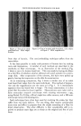
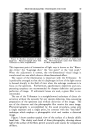
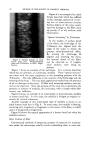
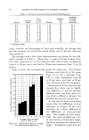
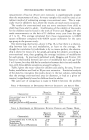
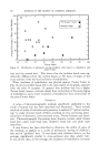
82 JOURNAL OF THE SOCIETY OF COSMETIC CHEMISTS 20, 25, 30, 40 and 50 minutes of ultraviolet irradiation. This was done to ddtermine the erythema dose of ultraviolet light. Following this procedure three areas were examined with a reflection meter a control untreated area, the area exposed to 15 minutes of ultra- violet and the area exposed to 50 minutes of ultraviolet light. RESULTS REaCT•O• TO ULTRaV•OLEX L•C•T Left thigh, Right thigh, Time, min. ultraviolet alone 8-MOP plus ultraviolet 15 No erythema No erythema 20 No erythema No erythema 25 Faint erythema No erythema 30 Definite erythema No erythema 40 Definite erythema No erythema 50 Definite erythema No erythema The erythema dose of ultraviolet radiation on the left thigh was 25-30 minutes. Fifty minutes of ultraviolet radiation on the right thigh pro- duced no erythema. REVLECT•O• METER REim•C WroTE D•sc = 50 Left Thigh Right Thigh Untreated area 33 31 Area exposed to 15 min. ultraviolet 30.5 26 Area exposed to 50 min. ultraviolet 27 28 (erythema) The relationship between the control areas and those exposed to 15 minutes of ultraviolet radiation indicates greater absorption of light in the area exposed after the ingestion of 8-MOP (probably due to pigment formation). The absorption of the 50-minute area of the left thigh is due to erythema. The lack of increased absorption of light by the same area of the right thigh confirms the clinical impression that there was no ery- thema. MICROSCOPIC EXAMINATION Specimens of skin were removed with a cutaneous punch, fixed in 10 per cent formalin solution and stained with hematoxylin and eosin and sil- ver nitrate. SPECIMENS FROM THE LEFT THIGH These showed the well-known changes which follow ultraviolet irradia- tion. There was thickening of the horny layer and a minimal inflamma- tory infiltrate in the upper curls. The changes were more marked in the
Purchased for the exclusive use of nofirst nolast (unknown)
From: SCC Media Library & Resource Center (library.scconline.org)