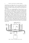
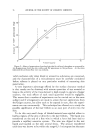
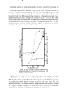
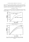
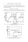
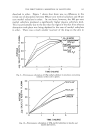
JOURNAL OF THE SOCIETY OF COSMETIC CHEMISTS his own words. We are fortunate that he is here in person to present his observations. Special histochemical methods can be used on a histologic section for detecting the presence of a substance which had been placed on the cu- taneous surface before the section was prepared (7). If care is used, this method can show whether or not penetration has occurred, but it will not give quantitative data. UsF. oF T•t^cF. Rs One of the early methods used for studying penetration of fats into skin employed an oil-soluble dye to color the fat globules. Histologic sections of the treated skin were then examined for the presence of dyed globules (8). When using any method in which a tracer is employed, one must always be suspicious that the tracer may become separated from the substance to which it was originally added and attach itself to some other substance. When a dyed fat globule is observed in a histologic section of skin, one must determine whether it is the globule to which the dye was originally attached or whether the dye has become dissolved in one of the fats previously present in the skin. If the dye has shifted from one fat to another, the presence of colored globules in the histological preparation is positive evidence only of penetration of the dye and provides no information about the penetration of the original fat. In a modification of the dye method, a fluorescent material rather than a dye is used, and the histologic sections are examined under a micro- scope, the condenser system of which can transmit ultraviolet radiation. This method is open to the same criticism as the one in which a dye is used as a tracer. One cannot be sure that the fluorescent material remains with the substance whose penetration is being studied. This objection is not valid if the penetrating substance is itself fluorescent. The penetration of vitamin A (9) and some hydrocarbons, e.g., benzyprene (10), which are fluorescent, has been studied by means of fluorescent microscopy. One pitfall in this method is the fact that skin naturally contains substances which fluoresce at a wavelength quite similar to that of the fluorescence from vitamin A and benzpyrene. Fluorescence from normal constituents of the skin must be differentiated from that emanating from absorbed substances. In recent years, the use of radioisotopes for tracing penetration (11) has become increasingly popular. As a rule, an attempt is made actuall.y to synthesize the substance being studied so that it includes a radioisotope. At times, the molecular structure of the penetrating substance is modified somewhat by incorporation of the radioactive element, as for instance in the iodination of an unsaturated fat with I TM (12). In working with radioactive compounds, one must always make certain that the radioactive

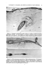
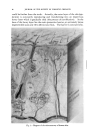
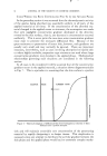
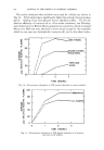
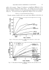
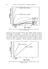
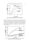
STATEMENT OF PROBLEM AND CRITICAL REVIEW OF PAST METHODS 63 molecule remains unchanged as it penetrates into the skin. Otherwise, one could not be certain that the presence of radioactivity signified any- thing more than the presence of the radioisotope. In a material like an iodinated fat, the iodine might split from the fat. In our own laboratories, we have observed that a portion of an anticholinesterase agent, synthesized with radioactive phosphorous, undergoes decomposition while passing into the skin. Therefore, unadjusted measurements of the total radio- activity in the skin following exposure to this substance would be misleading and would indicate that the amount of anticholinesterase agent present is greater than it actually is (13). The surfactant, dodecyl sulfate (syn- thesized with SaS), on the other hand, appears to penetrate unchanged, and a direct measurement of radioactivity following penetration of this substance would be a true measure of the amount of dodecyl sulfate absorbed (14). Radioactive tracers have been used for the purpose of learning the pathways of penetration. Autoradiography is a useful technique, but it also has some limitations and pitfalls. One must remember that an autoradiogram shows the position in the skin of a radioactive compound at the time the autoradiogram was made it does not necessarily show the pathway by which the radioactive substance reached that position. Evidence of radioactivity in the hair follicle and sebaceous gland following application of a radioactive substance to the cutaneous surface does not necessarily signify that the substance has reached the gland via the hair follicle. An autoradiogram of a section of skin, prepared after surface contact with radioactive sodium laurate, in which the densest shadow corresponds to the erector pili muscle could be shown. Certainly, you would readily agree that it would be absurd to say that such an autoradiogram indicates that the route of penetration has been via the erector pill muscle. Only a series of autoradiograms taken at intervals following the application of a radioactive substance to the cutaneous surface will show the progression of that substance through various pathways in the skin. For those of you who consider work with autoradiography, but have had no previous experience with it, I should like to refer you to the textbook by Boyd (15) on this subject and to warn you against three pitfalls: first, when a radioactive element which emits high-energy radiation is being used, there will be considerable scatter on the autoradiogram. That is, the palticle emitted from the radioisotope may travel some distance from the site of the isotope before striking a silver granule and producing a shadow. Therefore the position of the shadow does not always correspond to the ex- act position of the radioactive compound. Second, if the tissue is being placed directly on the photosensitive emulsion, reducing substances in the tissue may react chemically with the
Purchased for the exclusive use of nofirst nolast (unknown)
From: SCC Media Library & Resource Center (library.scconline.org)