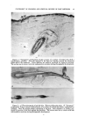
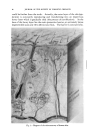
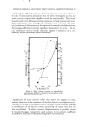
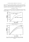
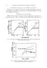
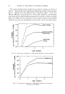
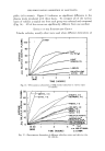
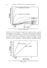
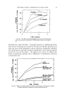
STATEMENT OF PROBLEM AND CRITICAL REVIEW OF PAST METHODS 63 molecule remains unchanged as it penetrates into the skin. Otherwise, one could not be certain that the presence of radioactivity signified any- thing more than the presence of the radioisotope. In a material like an iodinated fat, the iodine might split from the fat. In our own laboratories, we have observed that a portion of an anticholinesterase agent, synthesized with radioactive phosphorous, undergoes decomposition while passing into the skin. Therefore, unadjusted measurements of the total radio- activity in the skin following exposure to this substance would be misleading and would indicate that the amount of anticholinesterase agent present is greater than it actually is (13). The surfactant, dodecyl sulfate (syn- thesized with SaS), on the other hand, appears to penetrate unchanged, and a direct measurement of radioactivity following penetration of this substance would be a true measure of the amount of dodecyl sulfate absorbed (14). Radioactive tracers have been used for the purpose of learning the pathways of penetration. Autoradiography is a useful technique, but it also has some limitations and pitfalls. One must remember that an autoradiogram shows the position in the skin of a radioactive compound at the time the autoradiogram was made it does not necessarily show the pathway by which the radioactive substance reached that position. Evidence of radioactivity in the hair follicle and sebaceous gland following application of a radioactive substance to the cutaneous surface does not necessarily signify that the substance has reached the gland via the hair follicle. An autoradiogram of a section of skin, prepared after surface contact with radioactive sodium laurate, in which the densest shadow corresponds to the erector pili muscle could be shown. Certainly, you would readily agree that it would be absurd to say that such an autoradiogram indicates that the route of penetration has been via the erector pill muscle. Only a series of autoradiograms taken at intervals following the application of a radioactive substance to the cutaneous surface will show the progression of that substance through various pathways in the skin. For those of you who consider work with autoradiography, but have had no previous experience with it, I should like to refer you to the textbook by Boyd (15) on this subject and to warn you against three pitfalls: first, when a radioactive element which emits high-energy radiation is being used, there will be considerable scatter on the autoradiogram. That is, the palticle emitted from the radioisotope may travel some distance from the site of the isotope before striking a silver granule and producing a shadow. Therefore the position of the shadow does not always correspond to the ex- act position of the radioactive compound. Second, if the tissue is being placed directly on the photosensitive emulsion, reducing substances in the tissue may react chemically with the

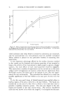
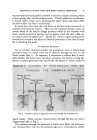
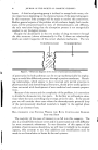
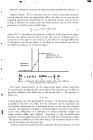
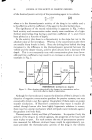
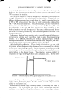
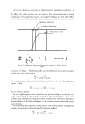
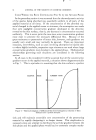
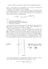
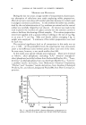
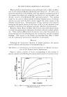
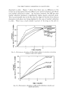
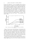
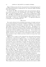
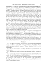
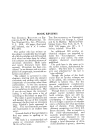
64 JOURNAL OF THE SOCIETY OF COSMETIC CHEMISTS emulsion and produce a shadow even when no radioactivity is present. Such shadows in the epidermis, follicular lining and around the sebaceous gland can be seen in Fig. 1, an "autogram" which was prepared from normal human skin containing no radioisotope. Third, if the tissue and photosensitive emulsion are on separate slides which, during exposure, are clamped tightly together, a pressure or stress artifact can be produced from hard tissues when no radioactivity is present. Figure 2 is an "autogram" in which the shadow was produced by pressure of the hair against the emulsion no radioisotope was present. MEASUREMENT or PHYSIOLOGICAL REACTIONS Development of a wheal following application of histamine to the skin (16), vasodilatation following application of nicotinic acid esters or other vasodilators (17), and anesthesia following topical application of local anesthetics (18) are examples of physiological reactions which have been used in studying the penetration of pharmacologically active substances. Reactions of this sort give a clear yes-or-no answer to the question of penetration: if a reaction is produced, the penetrating substance must have reached the deeper layers of the skin where lie the blood vessels and nerves. Such reactions, however, may not be directly proportional to the amount of material which has penetrated and seldom have methods of this kind yielded accurate, quantitative measurements of material which has penetrated. ANALYSIS or TISSUES The amount of a substance which penetrates into and through the skin after being applied to the cutaneous surface can be determined by analysis either of the skin itself, or, if there has been systemic dissemination, of some distant tissue. Before analyzing any specimen of skin, it is of course essential that the cutaneous surface be thoroughly cleansed of any residue of the applied material which has not penetrated into the actual tissue of the skin. With some materials, this is difficult to accomplish. If one is observing penetration into the skin only, then this method is applicable to either excised skin or skin on the living animal. Analysis of full-thickness skin provides no indication of how far into the skin the applied substance has penetrated. The results of analysis would be the same, whether the applied substance was only in the stratum corneum or had penetrated below the major barrier. If a liquid receptor is held in contact with the derreal side of a specimen of thin, excised skin, it is sometimes possible, by analyzing the receptor fluid, to determine the amount of applied material which has passed entirely through the skin (19). By analysing the receptor fluid at successive intervals, the rate of penetration may be determined by a test of this type.
Purchased for the exclusive use of nofirst nolast (unknown)
From: SCC Media Library & Resource Center (library.scconline.org)