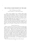
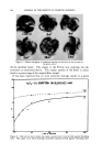
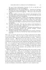
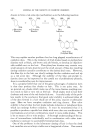
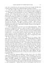
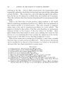
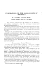
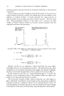
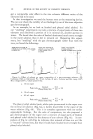
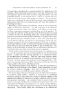
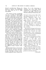
THE NATURAL COLOR PIGMENTS OF THE HAIR By S. W. BEcKE, JR., M.D.* Presented September/5-/6,/.960, Seminar, Chicago LOWER ANIMALS, regardless of hue or brilliance, appear content with the outer covering supplied by nature. The externally unsatisfied human being insists upon alterations. While our society invokes the aid of brown body paint, multicolored lip grease and nail lacquer as well as eyelid, lash and brow coloring, its main efforts are directed at the hair. Tinted hair is utilized to attract attention, conform to social fad or as part of the dogged struggle against age. Complete control of hair color ne- cessitates understanding of the natural hair pigments. •iln spite of the many outstanding investigators who have probed the problems of hair pigment there are few universally accepted facts. In many areas we have only floating hypotheses firmly attached to vague conjecture. This paper will attempt to outline the prevailing winds of present opinion with emphasis on findings related to the control of hair color. PIGMENT FORMING CELLS Pigment in human hair is produced in the melanocytes, dendritic cells, lying in the upper hair bulb at the superior border of the dermal papilla (Fig. 1). These cells migrate from the neural crest into the skin during early fetal life. (1). They are carried to their final position by the de- veloping hair germ. Although the melanocytes of the hair tend to be larger than those of the skin they are embryologically identical. While the cell body lies at the edge of the derreal papilla the dendritic processes grow upwards insinuating themselves between the undifferentiated cells of the cortex and medulla. The method by which pigment leaves the dendrites is not completely known and may be variable. It appears that portions or entire dendrites are broken off and engulfed by or carried be- tween the cells of the hair as they grow upward (2). The melanocytes re- place themselves by mitotic division. A few melanocytes remain in si•u when the hair is shed and these give rise to the pigment forming cells of the new hair. * Dept. of Dermatology, College of Medicine, University of Illinois, Chicago, Ill. 127

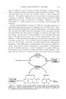
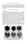
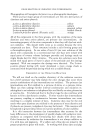
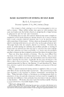
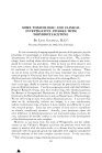
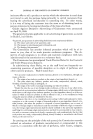
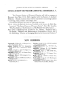
128 JOURNAL OF THE SOCIETY OF COSMETIC CHEMISTS PIGMENT GRANULE FORMATION Observations with the electron microscope by Barnicot and Birbeck (3, 4) appear to provide the best description of this phenomenon. Pigment granules appear to arise in the Golgi zone of the melanocytes. Vesicles in the Golgi zone increase in size and a structure composed of concentric membranes or a crumpled spiral membrane forms in this space (prepigment granule). This structure is presumed to be protein. In the dark hair, pigment is deposited within this protein matrix until the granule becomes a dark homogenous structure. The granules in blond hair are fewer and less well developed but appear to be the same type. Granules in red hair, however, are smaller, have an ill-defined boundary and contain groups of small dense particles. After the granules form in the Golgi zone they become evenly dispersed in the cytoplasm. The granules eventually move into the dendrites and exit from the melanocyte. It is presumed that the enzyme governing pigment formation becomes active on the prepigment granule before pigment is formed. The electron micro- scope provides no evidence regarding exact location of the enzyme. The mechanism by which pigment attaches to the protein matrix is unknown. Hair melanocytes in the albino produce granules consisting only of protein matrix. These granules disperse through the cytoplasm but cannot be followed into the hair. Gray hair from one individual was studied. The melanocytes could not be identified and no structures resembling pigment granules were found. Pigment granules persisted as identifiable structures in dark hair which had been bleached with peroxide. ENZYMES AND PIGMENTS The basic enzyme of human pigment formation is tyrosinase (5, 6). This enzyme is a copper protein which is found only in the melanocytes. It Figure 1• Nor•ai iu•an black hair. A. Melanocytes occupy upper hair matrix. I-Ien•a- .toxylin and eosin. B. Radioautographic tyrosinase method. Lithium carmine. Tyrosinase m the hair bulb melanocytes had catalyzed the oxidation of 0 4 labeled tyrosine to 0 4 labeled melanin. The sites of radioactive melanin are represented by the dense masses of silver grains. (Figure originally published in The Biology of Hair Growth, Montagna, W., and Ellis, R. A., editors, "The Nature of Hair Pigment," by Fitzpatrick, T. B., Brunet, P., and Kukita, A. Academic Press, Inc., New York, p. 288, 1958.)
Purchased for the exclusive use of nofirst nolast (unknown)
From: SCC Media Library & Resource Center (library.scconline.org)