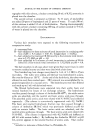
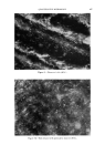
QUANTITATIVE MICROSCOPY 501 Figure 14.--Hardness test on nail polish (interference micro- scope: 200)). The lower limit of the measuring range of interference microscopes be- comes important in such applications of the cosmetics industry which deal with the pharmaceutical and cytological testing of materials, where de- terminations of dry and wet mass are made and concentration measure- ments are of interest. Most of these will be carried out in histological sections or in smear preparations. It may be interesting to calculate the limits of the method and the sensitivity of detection to evaluate the method for work in penetration studies. To reach the lower limit of interference methods one must employ micro- densitometric methods and use the interference contrast adjustment of the instrument. Only one interference fringe is spread out over the field of view. It's intensity distribution is recorded. The optic path introduced by the specimen causes it to appear darker than its surroundings. Loca- tions of equal intensity--or equal density on the photographic plate-- correspond to equal optical path, so that the path differences introduced by the specimen can be determined directly by a microdensitometric measure- ment. As a rule, a phase shift of approximately •/•00 of a wavelength (about 20 A.) is the limit of detectable specimen influence. A better idea of the sensitivity of the method is obtained when one ex- presses this measurable optic path difference in terms of detectable changes in dry mass or concentration. A 20 A. path difference in a histological section of 5u thickness corresponds to a change in refractive index of P/t = n, -- n = 2 m/z/5000 m/z = 4 ) 10 -a

502 JOURNAL OF THE SOCIETY OF COSMETIC CHEMISTS To convert this value into differences of concentration of a substance, one has to know the specific refractive increment, i.e., the increase of the refractive index of a solution for every 1% increase in concentration. For most biological materials, such as proteins, carbohydrates or lipids, this value is 1.85 X 10 -3 (13). Using this value, it follows that an in- crease in concentration of about 0.2% could be detected. In order to ob- tain practical numerical values for the specific refractive increment the concentration is expressed in g./100 cm. 3. The volumes of biological cells or cell components are very small. A volume of 15• 3 shall be assumed. Since a change of 200 rag. in 100 cm. 3 is detectable, in a volume of 15• 3 this amounts to only 3 X 10 -ng. In this application, the interference microscope thus becomes a highly sensitive optical balance. Such a highly sensitive method is not only applicable to biological prob- lems but also offers opportunities in the study of dissolving rates, of the effect of protective colloids added to soaps and lathers, and interactions between detergents and films of a fatty nature. Interference microscopical methods thus offer a possibility to measure quantitatively those effects of a cosmetic treatment or preparation and of colloid chemical processes which produce a change in optical path. For practical purposes, measurements can detect changes of optical path from around 1 min. to 20 A., a range of approximately six orders of magnitude. ANALYSIS IN POLARIZED LIGHT One of the most revealing microscopic methods is the examination of microscopic structures in polarized light. Numerous materials are either crystalline, as, for example, many organic substances, or they have sub- microscopic crystalline regions, for instance, gels (23, 24). Such structural anisometry leads to optical anisotropy. The anisotropy characteristics of such specimens can very accurately be measured in a polarizing microscope by analyzing the state of polarization of light which has passed through them. Even the molecular arrangements in the structures of these mate- rials can be derived from such studies. Analysis in polarized light can lead to highly interesting information on the orientation of submicroscopic elements, micelles and gel components in anisotropic materials. On the other hand, polarized light can also serve as a highly sensitive method of detection. In fact, in some specialized polarizing microscopes with rectified optics, optic path differences of as little as 0.1 A. have been detected (25). But even though the sensitivity of standard polarizing microscopes will not go quite as high, it is more than adequate for most purposes. The full measuring range of a 1/30 wavelength mica compensa- tor covers 170 A., and its lower sensitivity limit is of the order of magnitude
Purchased for the exclusive use of nofirst nolast (unknown)
From: SCC Media Library & Resource Center (library.scconline.org)