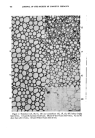
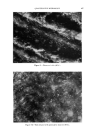
TOPICAL PREPARATIONS AND THE HEALING OF SKIN WOUNDS 511 lP'ounding Procedures On the day prior to inflicting the wounds, the hair on the backs of the animals was removed with an electric clipper. With the animals under ether anesthesia, an incision approximately 25 mm. in length was made along the midline of the back, starting at the shoulders and proceeding caudally. In order to cut only the skin and not involve underlying tissue, an initial small slit was made in the skin with a sharp, fine pointed scissors. The skin was then raised by grasping it with forceps at a point just an- terior to the slit and a scapel blade, cutting edge upward, was inserted into the opening and drawn through the skin to a point about 25 min. from the point of origin. The edges of the wound were approximated and then closed with five equally spaced interrupted sutures (40 gauge stainless steel wire). Treatment was begun immediately. On the sixth day after the wound was inflicted, the sutures were removed. Tensile Strength Measurements The mice were sacrificed and hair that had regrown was carefully clipped with a small scissors. A wide section of skin containing the healing wound was excised. Care was taken to include only the skin and closely adhering subcutaneous tissue. The skin section was then placed on a plate-glass block that was moistened with normal saline. This permitted the skin to assume what might be termed a standard dimension. A strip of skin along the posterior (caudal) end of the wound, perpendicular to the line of the wound, was trimmed off and discarded. Then a strip 8 mm. wide was cut off and used for the tensile strength measurements. The next 2-3 mm. section of the wound was removed and fixed in formalin for histological examination. Another 8 min. segment was then obtained from the re- maining (anterior) portion of the wound, for a second tensile strength de- termination. A modification of the Charney, Williamson, Bernhard (42) technique was used to determine the tensile strength of the healing wound segments. One end of the skin section was mounted in a clamp, and a lightweight basket lined with a polyethylene bag was attached to the other end. With the skin segment in a vertical position, weight in the form of dry, free flowing sand at a rate of 7 g./sec. was added to the basket until rupture of the wound occurred. The results are expressed as grams or weight required to separate the edges of the healing wound. Averages for each group were calculated and include the values obtained with both segments of wound from each mouse. Four series of tests were conducted. The first was for the purpose of checking the basic method and to construct a time rs. wound tensile strength curve, with the animals under control conditions, i.e., no treat- ments applied to the wounds. Following this the experiment was re-

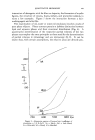
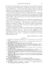
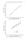
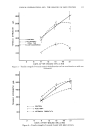
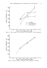
512 JOURNAL OF THE SOCIETY OF COSMETIC CHEMISTS peated using available materials which had been reported to have an ef- fect upon wound healing. The following preparations were evaluated: Series Treatment Material II (A) Ointment containing Vitamins A and D (B) Liquid containing 2% allantoin and 5% refined coat tar extract (C) Cream containing 0.01% 6%9a-ditquoro-16a-hydroxy- prednisolone acetonide III (A) /lloe vera extract in an ointment base (B) Essential oil (steam distillate) from leaves of.4rtemisia tridentata IV (A) Ointment containing 0.01% 6a,9r•-difluoro-16a-hydroxy prednisolone acetonide (B) Cream containing 2% pantothenylol All test materials were applied once daily over the healing wound and surrounding skin until the day on which the tensile strength measurements were made. Series I. Control Group Sixty mice were divided into six groups of ten animals each. After surgery, each animal was housed in an individual cage and given food and water ad libitum. The tensile strength of the healing wounds was de- termined 6, 9, 12, 15, 18 and 21 days after wounding, with one group of mice being sacrificed at each time interval. Figure 1 shows the results obtained over the 21 days experimental period. There is a time-related increase in the tensile strength of the wounds. The ranges indicated above and below the central point at each interval represent the standard error calculated for that group of measure- ments. Through the 15 day determinations, the standard errors remained fairly constant at about plus or minus 20 g. At 18 and 21 days after sur- gery a wider range was observed as higher tensile strengths were reached and the standard errors increased to 40 to 50 g. A simple calculation showed that despite the absolute increases in standard error, these values generally represented between 5 and 10% of the group average. Results obtained indicate that with the basic techniques employed statistically acceptable measurements could be made and that the determination of increases in tensile strength may be a useful indication of the skin wound repair process. Series H Ninety-six mice were used in these tests. They were divided into six- teen groups of six animals each and wounded and housed as in Series I. Five groups served as controls, and five groups received daily applications
Purchased for the exclusive use of nofirst nolast (unknown)
From: SCC Media Library & Resource Center (library.scconline.org)