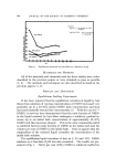
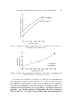
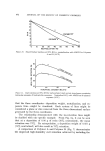
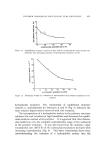
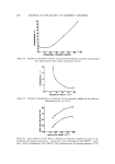
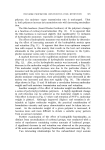
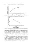
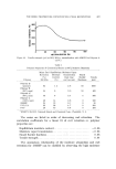
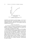
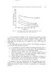
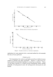
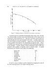
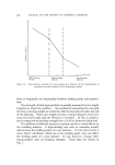
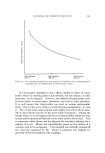
838 JOURNAL OF THE SOCIETY OF COSMETIC CHEMISTS especially in the female, who undergoes a rather abrupt decline in hormonal output at the menopause, the epidermal sweat and sebaceous glands become much less active, almost to the point of degeneration. Although the exact method of action of the sex hormone on the cell layers of the skin is not known, it is recognized that the "female" hormones and hormone-like steroids, when topically applied to aged skin, effect a histological picture in the epidermis which is strikingly similar to that of a young person. One area of investigation concerning the epidermis and aging that has been studied to some degree is that of rate of turnover of the cells of the epidermis. The turnover time, or renewal time, is usually defined as the average time for all the cells in the basal layer of the epidermis to reach the horny layer. Although several studies have been undertaken to measure this turnover time, various sources have been used for epidermal sections, and many different areas of the body have been used for tissue sampling. This lack of coordination between donor and site, coupled with infrequent recording of the donor's age, has unfortunately led to an unclear presentation of the relation of epidermal turnover to aging. Weinstein (25) has deduced values for turnover time using skin from the back of an adult pig. The morphology of the pig epidermis has been found to be strikingly similar to human epidermis. He found a transit time of 14 days through the viable cell layer, and 16 days through the horny layer, giving a total turnover time of 30 days. In working with the rat, Storey and Le Blond (26) reported a turnover time of 19 days in plantar epidermis taken from an adult animal. Bertalanffy (27), also using adult rats, found that the renewal time for the abdominal epidermis was 19 days. In a second study, Bertalanffy et al. (28) com- pared epidermal growth in young, adult, and senile rats. They found that when the total thickness of the epidermis from different age groups was compared, the senile skin showed a reduction by about two to three layers in the epidermis taken from ear or abdomen. In areas such as the bottom of the foot, there was an increase by one or two in the number of cell layers. As regards the mitotic rates, it was found that there was a general increase by about 50% in the rate of turnover of the senile epidermis as opposed to the young. In a similar study using human abdominal skin samples, Thuringer and Katzberg (29) observed an increase in the mitotic index such that the epidermis of the senile individuals was dividing twice as fast as that of the young group. The cause of this increased epidermal turnover is not yet positively

CHANGES IN HUMAN SKIN WITH AGING 839 known. However, one might be tempted to postulate a feedback con- trol mechanism operative in the epidermis. As was mentioned, the adult and senile skin show increased desquamation, possibly because of increased keratinization, modification of dermal vascularity, or insuffi- ciency of epidermal glandular secretion. This cell loss may act as a trigger to signal an increase in the replacement of cells. As cell loss is accelerated, then, so also is cell replacement. While there are but few techniques presently available for the treatment of aging skin, the ultimate method will probably involve the use of agents which will be able to act directly on the controlling mech- anisms of the body's cells--the genes--either to correct errors (which, as we have seen, can evolve with time), or to initiate some desired ac- tivity (such as induction of a specific enzyme). When man is able to achieve this kind of control, there will be practically no limit to his ability to regulate the functioning of his body even to stemming the tide of aging. (Received April 17, 1968) REFERENCES (1) Johnson, H. D., Kintner, L. D., and Kibler, H. H., Effects of 48øF and 83øF on lon- gevity and pathology of male rats, J. Gerontol., 18, 29 (1961). (2) McCay, C. M., Magnard, L. A., Sperling, H., and Barnes, L. L., Retarded growth, lifespan, ultimate body size and age changes in the albino rat after feeding diets re- stricted in calories, J. Nutr., 18, 1 (1939). (3) Curtis, H. J., Biological mechanisms underlying the aging process, Science, 141, 686 (1963). (4) Albert, M.D., X-irradiation induced mitotic abnormalities in mouse liver regenerating after CC14 injury. I. Total body irradiation. J. Nat. Cancer Inst., 20, 309 (1958). (5) Stevenson, K. G., and Curtis, H. J., Chromosome aberrations in irradiated and nitrogen mustard treated mice, Radiation Res., 15, 774 (1961). (6) Bjorksten, J., Chemical causes of the aging process, Proc. Sci. Sect. Toilet Goo&, Assoc., 41, 32 (1964). (7) Verzar, F., The aging of collagen, $ci. Am., 208, 104 (1963). (8) Harman, D., The free radical theory of aging: effect of age on serum copper levels, J. Gerontol., 20, 151 (1965). (9) Norins, A. L., Free radical formation in the skin following exposure to ultraviolet light, J. Invest. Dermatol., 39, 445 (1962). (10) Lorincz, A. L., Physiology of the aging skin, [llinois Med. J., 117, 59 (1960). (11) Sams, W. M., Jr., and Smith, J. G., Jr., Alterations in human dermal fibrous connective tissue with age and chronic sun .damage, in Montagna, W., Advances in Biology of the Skin--Volume VY--Aging, Pergamon Press, New York, 1965, pp. 199-210. (12) Rothman, S., Water and electrolytes, in Physiology and Biochemistry of the Skin, The University of Chicago Press, Chicago, 1961, pp. 493-514. (13) Flesch, P., The chemistry of the aging skin, J. Soc. Cosmetic Chemists, 6, 377 (1955). (14) Smith, J. G., Jr., and Finlayson, G. R., Dermal connective tissue alterations with age and chronic sun damage, Ibid., 16, 527 (1965).
Purchased for the exclusive use of nofirst nolast (unknown)
From: SCC Media Library & Resource Center (library.scconline.org)