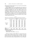
ADHESIVE-TAPE STRIPPING TECHNIQUE FOR EPIDERMAL HISTOLOGY 4•33 Neck is apparently intermediate between face and trunk. Wolf put it with face. We put it with trunk, but it does exhibit one of the characteristic features of cheek and forehead, i.e. nucleated cells. Our classification relates fairly well with patterns of use and exposure. The surfaces of the trunk, arms, and legs are mostly covered and unstressed. Palm and sole are an obviously unique pair of regions. Cheek and forehead are continuous- ly exposed to the environment. Neck is partly so exposed. Back of hand and top of foot also get partial exposure to environmental insults or to occlusion and friction, both of which seem to produce a similar surface corneum pattern. The back of the hand can be subjected to severe insult by the normal vicissitudes of weather, work and washing this may be reflected in the fact that nucleated corneum cells can sometimes be obtained from the back of the hand. This regional variation also involves corneum cell size. Wolf (2) found cells of back of hand smaller than those of scrotum which were smaller than those of stomach. We have confirmed the size relationship between back of hand and stomach, and can add that palm and forehead corneum cells are smaller than those of back of hand, and cheek cells are even smaller. CONCLUSIONS We have developed and tested by frequent use over several years, a quick, easy, and reliable tape-stripping method for investigating the struc- ture of the skin's surface layers. It involves application of Sellotape to skin with a roller, and transference of cells to albumin-coated slides. This technique has enabled us to recognise three distinct stages in the maturation of corneum cells. The same pattern of distribution of those cell types was found in mouse, rat, hamster, guinea pig, rabbit, and man. There are differences in the ease with which the corneum of different species can be stripped. There may also be some differences in corneum cell diameter between species. The diameter of corneum cells of 4-day old rats was smaller than that of 2 to 3..week old rats. In human corneum regional variation was recognisable. Four groups of regions could be distinguished by the patterning and grouping of their surface cells: trunk and limbs, dorsum of hand and foot, palm and sole, cheek and forehead. Some of those regions also differed in corneum cell diameter. (Received: 9th September 1968)

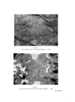
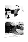
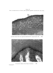
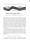
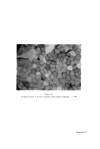
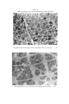
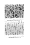
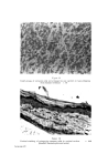
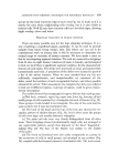
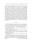
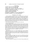
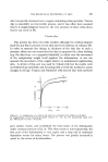
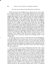
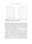
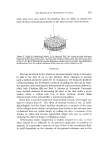
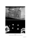
464 JOURNAL OF THE SOCIETY OF COSMETIC CHEMISTS REFERENCES (1) Pinkus, H. Giorn. Ital. Dermatol., 107, 1115 (1966). (2) Wolf, J. Z. Mikr. Anat. Fosrch., 46, 170 (1939). (3) Wolf, J. Z. Mikr. Anat. Forsob., 47, 351 (1940). (4) Keddie, F., Orr, A. and Liebes, D. Sabouraudia, 1, 108 (1961). (5) Goldschmidt, H. and Kligman, A.M. Arch. Dermatol., 06, 572 (1967). (6) Brody, J. J. Ultrastruer. Res., 4, 264 (1960). (7) Brody, J. Acta Dermato-Venereol., 40, 74 (1960). (8) Snell, R. Z. Zellforsch., 70, 492 (1967). (9) Hope, J. Personal communication (1968). (10) Christophers, E and Kligman, A M. J. Invest. Dermatol., 4•,, 407 (1964). (11) Epstein, W. L. and Maibach, H. I. Arch. Dermatol., 0% 462 {1965). (12• Porter, D. and Shuster, S. J. Invest. Dermatol., 40, 251 (1967). Introduction by Mr. Jenkins I wish to emphasize two important points on the method. Firsfly, in order to get the best possible tape strippings, one must ensure the optimum contact between the tape and the skin. This is why, whenever possible, we excise the skin, mount it on a smooth, firm support, and stretch it slightly to remove surface wrinkles. It is not always convenient to excise skin, but when it can be done, it is advisable to do so. Secondly, one of the most important requirements for getting good transfer of cells from the tape to the slide is to ensure that the cells are firmly stuck to the slide before starting to dissolve the adhesive of the tape. I also wish to provide additional information about corneum cells. Thickness of corneum strippings In many strippings there are relatively large areas where the cells are so trans- luscent that the layer is almost certainly only one cell thick. But in places the stripping is more opaque, abruptly so, with sharp demarcation at edges of cells. These darker groups of cells are probably more than one layer thick. The second, and related, point is that in the thicker areas the cells appear to be neatly superimposed on the ones beneath they do not overlap half-way across their subjacent neighbours (Fig. 17). Since we frequently see this type of arrangement in tape strippings we are led to suppose that corneum cells tend to be arranged in vertical stacks. Clear-centred cells Their pale central areas are not merely shapeless spaces they have distinct boundaries which can be shown very clearly by careful staining. These clear centres are not always completely empty. They often contain a small, dense body which, one is tempted to presume, is the remains of the nucleus, even though it is not particularly basophilic (Fig. 18). Pycnotic nuclear cells These are situated more deeply in the corneum and usually look somewhat distorted in tape strippings, probably because they came from a depth where cells are soft, and consequently easy to distort. They are often rather more basophilic than more superficial corneum cells (Fig. 19). Nucleated human corneum cells These look essentially like corneum cells, but they have large, prominent, and
Purchased for the exclusive use of nofirst nolast (unknown)
From: SCC Media Library & Resource Center (library.scconline.org)