ADHESIVE-TAPE STRIPPING TECHNIQUE FOR EPIDERMAL HISTOLOGY 453 •-•ETHODOLOGY Our routine method is made up of the following stages: 1. Remove hair by plucking. 2. Mount excised skin on a wooden block 76mm X 12.5mm or 76mm X 25mm with weights hung on all sides. 3. Apply end of tape, roll tape on to skin with metal roller of appro- priate weight. 4. Remove tape by pulling it back quickly on itself, from posterior to anterior. 5. Fix stripping in formol-acetic-alcohol for 15 s. 6. Transfer to slide put tape on albuminised slide, smooth out, cover with blotting paper, apply enough pressure to keep tape flat. 7. Incubate at 60øC for 1 h. 8. Remove tape by putting slide into toluene for one to two days. After removal of tape put slide in toluene for another 2 h to remove all adhesive. 9. Stain cells e.g. in haematoxylin and eosin. 10. Dehydrate. 11. Clear. 12. Mount in DPX. 13. Dry preparation in incubator overnight. 14. Clean slide and label it. In the course of the development of this technique we examined all of the readily-available tapes. Some wrinkled badly in fixative, some fluoresced in uv light, some stretched, some stripped better than others, some allowed better transference than others, some left a stainable residue of adhesive on the slide. No tape was significantly better than Sellotape in overall suitability. Sellotape is very readily available, and we use it for routine work. Certain other tapes have special properties which may make them appro- priate for particular jobs. Hair can be removed from the skin in many ways. The best method is plucking o[ telogen hair. Clipping, with electric clippers, is preferable to wax depilation or shaving, which both alter the skin surface to an extent which is undesirable in this context. Stripping can be done more efficiently on excised skin put on a flat, firm surface than in its natural situation. Whenever possible we excise skin, and put it on a wooden block. Slight stretching is advisable to remove folds and wrinkles. Rat skin is adequately tensioned by 120 g hung on each

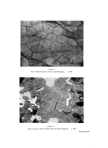
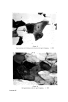
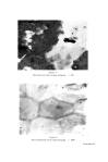
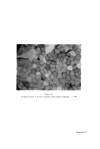
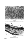
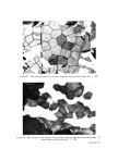
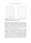
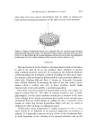
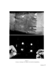
454 JOURNAL OF THE SOCIETY OF COSMETIC CHEMISTS side. The amount of tissue removed by the tape is to some extent propor- tional to the pressure with which tape is applied. A 12.Smm diameter stainless steel roller weighing 300 g gives good results with excised rat skin. When stripping intact skin we press the tape on with a wooden wall-paper roller, using moderate pressure. Stripping along the longitudinal axis is more efficient than transverse, on rats and mice. On hairy skins, stripping against the direction of hair growth is the better, that is, posterior to anterior. Fixation is probably not necessary for mature corneum cells, but is advisable if one is interested in immature epidermal cells. The Sellotape adhesive is dissolved by benzene, carbon tetrachloride, toluene, and xylene, and these all allow transference of cells to albuminised slides. Acetone, chloroform, and ether do not dissolve the Sellotape adhesive. APPEARANCE OF EPIDERMAL CELLS IN TAPE-STRIPPINGS Most of our work has been done on rats. We will therefore first des- cribe what we find in tape-strippings of rat skin. There are three quite distinct types of corneum cells. First of all the plain cell (Fig. 1) we call it plain because it has no remarkable features. It is just what everyone expects a corneum cell to look like. It is typically hexagonal and equilateral. In fact, it usually is hexagonal but not equi- lateral. It occasionally has five sides, and rarely four sides. There are no characteristic inclusions. Secondly, the cell with nuclear space (Fig. 2). This is basically like the plain cell, but has a lightly stained round or oval central area, with a fairly well defined boundary, and often containing a small dense body. The "space" looks like the space formerly occupied by the nucleus. We prefer not to call it a nuclear ghost. The term ghost suggests an attenuated replica of a nucleus. Thirdly, the cell with pycnotic nucleus (Fig. $). These are usually more distorted than the plain cells, but they are recognisably corneum cells. They are thin, flat, five or six sided, with no characteristic features except the presence of a small, dense, basophilic body resembling a pycnotic nucleus. This body is sometimes seen to lie in a lightly stained round or oval area. The granulosum cell (Fig. 4) is thin, flat, five or six sided, tending to be more rounded than corneum cells. It has a well-stained nucleus and the cell is full of rounded granules. The nuclear material is often separated into two or three or four pieces, sometimes of unequal size. Spinosum cells (Fig. 5) are easily distorted. They tend to be removed as
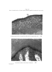
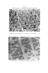
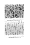
Purchased for the exclusive use of nofirst nolast (unknown)
From: SCC Media Library & Resource Center (library.scconline.org)