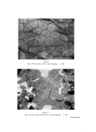
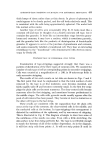
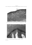
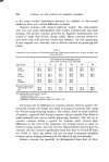
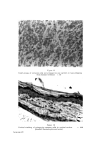
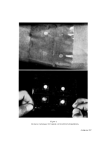
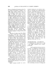
ADIIESIVE-TAPE STRIPPING TECHNIQUE FOR EPIDERMAL HISTOLOGY 465 healthy-looking nuclei. We have seen such cells in the top layers of corneum of human face, neck and back of hand, but not in strippings of human arms, legs, or trunk. They do not appear to be associated with any noticeable lesions (Fig. 20). Similar- looking cells have been seen infrequently in strippings from the flanks of rats and pigs. DISCUSSION MR. N. J. VA• ABB•: Have you been able to modify your tape stripping technique for use on the hairy scalp? MR. JE•:i•s: We have not attempted to take strippings from hairy scalp this is a fascinating problem I admit. M•. N.J. VA• ABBr,: Does your technique enable you to study micro-organisms in the epidermis more reliably than can be achieved by sectioning? MR. JE•Ki•s: Theoretically this ought to be a very profitable way of looking at their distribution. Difficulties might arise if one tries to transfer the skin cells and the micro-organisms on to slides. XYe know that the skin cells will transfer on to slides and can then be treated as fairly ordinary histological preparations whether the micro- organisms associated with the cells will transfer equally successfully, I just do not know. It is possible that the whole thing could be left on the tape then you merely have to decide whether you can stain the structures you want to see, without staining the adhesive and the tape backing and getting the whole picture obscured. For surveying a fairly large area this technique is very useful you can take a stripping 50 x 25mm quite easily and put this on the slide. If you can selectively stain the corneum cells you have some chance of estimating the depth you are getting down to. Tape stripping is variable in the thickness of the layers it takes off, but if you find cells with pycnotic nuclei, for example, in certain areas, you know you are pretty deep into the corneum. In this manner it should be very useful in each preparation you are seeing much more tissue than in a vertical section of skin. Dm D. SPRui•: How does the picture change under the influence of corticosteroids? The layers are then thinner and what is the change of the appearance of the cells? MR. JE•s: We have not done any work on the influence of that type of treat- ment. 1)•. N. A. I)u'r'rN•v•: Have you done any comparisons of strippings from the pahn of, say, an office worker compared with a building worker, and if so, what differences did you find? Mu. J•h'•s: So far we have not gone outside our ordinary environment, so •nost of our strippings do not give us any information on that type of variation. We have been using this techniqne for some time now, and know that it works very satisfactorily for certain purposes. We have started looking at the normal sources of variation--individual variation, and seasonal variation. I think the possibility of looking into variation in surface patterns caused by occupation and various other sources, should now be considered. DR. \¾. H•R'rs'ro•: After the eighth stripping with the biomicroscope you get a very luteresting picture. The surface, i.e. the remaining surface being left undisturbed,

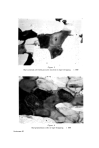
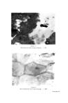
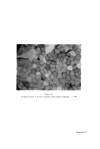
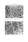
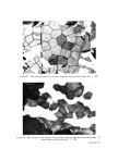
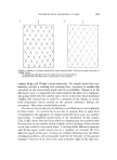
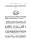
466 JOURNAL OF THE SOCIETY OF COSMETIC CHEMISTS shows a very curious situation in which the epithelial cells are stuck down except at their corners, and the corners tip up as though you had a deformed tile of which the four corners were pointing upwards. This is quite universal, and one gets the impres- sion that this is how the cells were lying, i.e that those cells that are deeply attached adhere over most of their centres and that their corners are free, and one begins to wonder whether there is a possibility of drug or fluid absorption from the surface through these looser, overlapping layers of corners of epithelial cells. MR. JENKINS: In tape stripping one frequently finds that cells deep in the corneum have a tremendous tendency to curl up, and it is in fact quite difficult to find fairly flat-looking cells in one's preparations which clearly show the characteristic features. I wonder whether the overlapping that appears to occur between the adjacent cells of human corneum means that when stripping is uneven, as it is especially in the deeper layers, you leave isolated cells sitting as islands, and their edges are free and might well curl up. The other possibility is that cells from deep in the corneum are presumably fairly moist and when exposed during tape stripping, there is apparently a tendency for such cells to dry out quickly and curl up. One has to be aware of that possibility of artefact, but vith human corneum cells the overlapping might well produce this type of curling up of the edges and corners that you have seen. DR. J. W. BOTHWELL: You showed some interesting pictures of stacked cells. Could you expand on that and indicate under what conditions or in what parts of the body you get the stacking, and whether regional or other varaiations cause a non- stacking type pattern? MR. JENKINS: We have not gone into this in any great detail. It is a very charac- teristic feature of vertical sections of guinea-pig skin one can almost spot a section of guinea-pig skin by this peculiar arrangement of corneum. Perhaps it is easily visible in the guinea-pig corneum because the layers become separated from each other in sectioning thus making this stacking visible. If one takes sections of rat dorsal comeurn and swells them in caustic (10) they frequently show small areas where very neat stacking of cells, one on top of the other, does occur. We have seen it in human tape strippings and, to a limited extent, in sections of human skin, but I could not say anything about the regional distribution of this stacking.
Purchased for the exclusive use of nofirst nolast (unknown)
From: SCC Media Library & Resource Center (library.scconline.org)