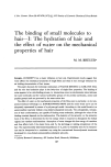
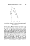
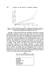
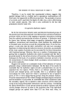
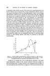
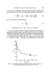
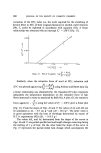
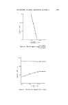
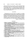
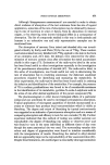
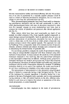
476 JOURNAL OF THE SOCIETY OF COSMETIC CHEMISTS concentrations of salt ions suppress the association of oppositely charged proteins (14) and Speakman and Whewell (15) have shown that wool fibres are much weaker in concentrated solutions of sodium chloride than would be predicted from the water activity of the solutions. This has been ex- plained (16) as being due to a reduction in thickness of the double layer around the --NHa + and --COO- groups in wool by the salt ions leading to reduced interaction between these groups. A similar role is envisaged for the salt ions considered in this study these penetrate the protein matrix and interact with oppositely charged and dipolar groups (e.g.--COO-, --NH• +, --OH, --NH•) located on the side chains of the constituent amino acids. Since such groups are responsible for the cohesiveness and mecha- nical strength of the corneum by forming hydrogen bonds and salt linkages between and within proteins when the salt ions are absent, the matrix is weakened by the presence of the ions. It is an established fact (10) that the hygroscopic substances which occur naturally in the stratum corneum cannot be removed by aqueous extraction alone. It is first necessary to remove lipids from the corneum by extraction with some suitable organic solvent after which water will leach the hygro- scopics from the structure. It would appear, however, that the simple ions used in this study could enter and be removed from the native corneum as well as from the extracted material without imparting any permanent damage to the structure. This was indicated by the return of the corneum strips to their original modulus values in water after thorough washing. The only conclusion that one can reach at this stage is that this is due to the difference in size between the small ions so far investigated and the larger naturally-occurring hygroscopic materials (organic acids, simple peptides etc). This observation is confirmed by the results obtained using a saturated sucrose solution for which the water activity is known. When native stratum corneum was equilibrated in this solution (Fig. 1) the modulus value ob- tained was identical to the vapour phase value at the same relative humidity (85•) indicating no additional affect due to the presence of the large sucrose molecules. In contrast, the modulus of the lipid-solvent extracted corneum was considerably less in the sucrose solution than in the vapour (Fig. 2). Clearly, removal of the lipid barrier had allowed penetration of the larger hygroscopi½ molecules (sucrose) with their resultant plasticizing effect on the corneum structure. An obvious difference, apart from size, exists between sucrose and the other species studied this is the fact that the sucrose molecule does not bear a charge. Since the absence of charge on the sucrose molecule should impart greater lipid solubility to it than

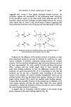
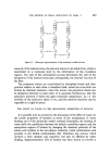
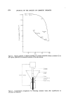
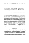
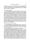
EFFECT OF SALT SOLUTIONS ON STRATUM CORNEUM 477 exists for the salt ions, then passage of sucrose through the lipid barrier should be facilitated for native stratum corneum if size was not the pre- dominant factor. It is suggested, therefore, that the ability of the corneum to retain its natural hygroscopic materials, when the lipid barrier is intact, is due, in some extent, to the size of the molecules which comprise the NMF. For reasons which have been discussed in the previous study (2) it has been suggested that the mechanical integrity of the corneum might reside in its intercellular junctions (junction = two adjacent cell membranes + desmosomes which act as intermediate adhesive material) rather than with- in the keratin filaments as had been assumed in the past (2). The present results, together with data obtained by Middleton (17) on the osmotic characteristics of the stratum corneum help to enhance this point of view. Middleton has shown that osmotic dehydration of stratum corneum occurs when it is bathed in a solution which has a higher osmotic pressure than the corneum cell contents. This will certainly be the case for the saturated solutions used in this study. Such a property can only be accounted for if significant amounts of the solutes do not penetrate into the corneum cells. If this is the case then the decrease in modulus values of the native corneum produced by the salt solutions in the present study must result from the action of these salts on the intercellular system furthermore, it is not too much of an extrapolation to suggest that the NMF is also located primarily in this region. It now only remains to consider the role of the lipids which allow pene- tration of small ions into the intercellular material but prevent the entry or removal of larger similar species. To this end, the salient features of a model for the junction between corneum cells are proposed in Fig. 4. The model consists of a protein/polysaccharide-rich central region (11) con- taining hygroscopic materials and isolated pools of lipid enclosed by the two adjacent cell membranes (lipid bilayers in association with proteins). The lipid bilayers of the cell membranes offer high resistance to the passage of charged species and water [complete hydration of the corneum requires •5 days (18)] and are considered to be responsible for the semi-permeable nature of the corneum. The large naturally-occurring hygroscopic molecules are retained within the central region of the junction by the lipid pools in the structure. That is to say, their removal by aqueous extraction is pre- vented due to steric factors. Extraction with lipid solvents removes all of the lipid and the hygroscopic substances are then readily removed with water, being leached out through the channels created by removal of the pools of lipid. The involvement of pools of lipid within the proteinaceous region of
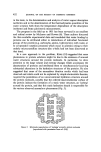
Purchased for the exclusive use of nofirst nolast (unknown)
From: SCC Media Library & Resource Center (library.scconline.org)