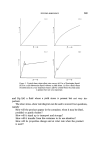
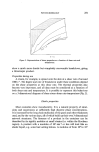
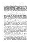
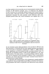
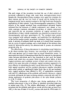
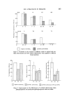
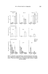
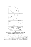
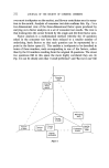
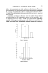
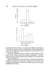
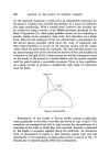
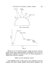
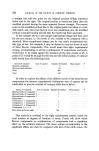
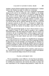
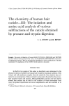
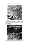
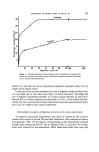
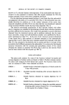
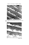
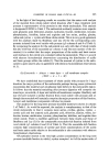
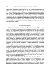
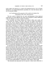
CHEMISTRY OF HUMAN HAIR CUTICLE--III 291 35- 30- 25- Pronase 3'0 4'0 5'0 6 ) 7 ) • 1•)0 Digestion time (h) Figure 1. Graph showing the gravimetric course of digestion of human hair cuticle with trypsin and with pronase. Each point shown represents the mean of two separate determinations. 260 about 115/o and that the slower dissolving component comprises about 9• by weight of the whole cuticle. In the case of the pronase treatment the rate of digestion within the first hour was very high and in that time some 22«5/0 of cuticle dissolved. Thereafter the rate of digestion diminished abruptly to a linear portion between 2« and 22« h. Beyond 30 h no further digestion occurred and in all 355/0 of the cuticle had dis- solved. The fast- and slow-dissolving components represent approximately 22«5/0 and 12«•o by weight of the cuticle respectively. Morphological progress of digestion of cuticle with trypsin and pronase An electron microscope examination was made of transverse hair sections treated with trypsin for 5 h and 70 h and then 'shadowed' with a mixture of carbon and platinum. After 5 h the regions corresponding to the endocuticle contained discrete holes measuring 0.05-0.1 gm in diameter (Fig. 2), and after 70 h larger holes were observed in the endocuticle. Often these latter holes were long and

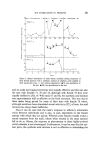
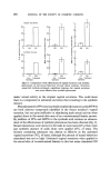
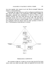
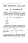
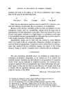
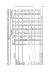
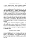
292 JOURNAL OF THE SOCIETY OF COSMETIC CHEMISTS narrow as if a discrete laminar subcomponent of the endocuticle had been dis- solved. It is also pertinent to mention that after trypsin treatment for 70 h the nuclear remnants of the cortex were completely dissolved. For the shadowed pronase-treated sections it was found that the endocuticle was peppered with holes at 2 h, and after 30 h (Fig. 3) the endocuticle was com- pletely dissolved. In addition at 2 h the nuclear remnants of the cortex were removed completely and after 30 h it was clear that the 'non-keratinous' material of the intermacrofibrillar matrix had also been degraded (Fig. 3). The electron microscope examination of hair sections stained with either silver or tungsten was valuable for determining in detail which cuticular structures had been affected by the enzymes. The results were generally in accord with those obtained from the shadowed sections and in addition indicated that the inner layer, exocuticle, A-layer and cell membrane complex were unaffected by the en- zyme treatments (cf. Figs 4-7). In the case of the extensive pronase treatments it was found that a thin network of material remained in the endocuticular regions (Figs 6 and 7). Since this latter material stained fairly intensely with silver (Fig. 7) it was assumed to be relatively rich in cystine and that it was part of the exocuticle which diffusely permeates the endocuticle. It is noteworthy that despite extensive treatments with the enzymes the intercellular cement of the cuticle (b-band) was stained as intensely with PTA (Figs 5 and 6) as the sections which had not been treated with the enzymes (cf. Ref. 2). Amino acid analyses The amino acid analyses for some of the fractions isolated by trypsin and pronase digestion of whole cuticle are listed in Table I. The first column contains the amino acid analysis of whole human hair cuticle. The fractions listed in the next six columns of Table I were obtained as follows: ENDO -- Soluble fraction obtained by digestion of whole cuticle for 5 days with pronase. EXO + A + I + M -- Insoluble material remaining after pronase digestion for 5 days. ENDO A- Soluble fraction obtained by trypsin digestion for 10 days. ENDO A1 -- Soluble fraction obtained by trypsin digestion for 9 h. ENDO A2- Soluble fraction obtained by taking the insoluble material remaining after trypsin treatment for 11 h and digesting it with trypsin for a further 10 days.
Purchased for the exclusive use of nofirst nolast (unknown)
From: SCC Media Library & Resource Center (library.scconline.org)