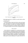
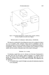
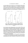
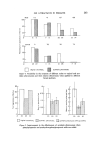
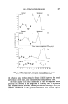
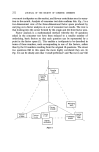
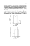
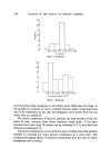
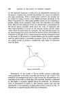
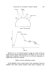
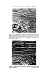
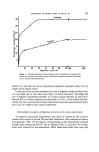
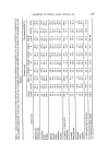
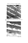
296 JOURNAL OF THE SOCIETY OF COSMETIC CHEMISTS the protein. Taking into account the fact that the cystine concentration of the cuticle cell membrane complex is quite low (2), we have calculated that the order of 1 amino acid residue in 3.7 is present as « cystine in the combined exocuticle. and A-layer. In our previous paper (2) we also presented evidence for suspecting that the cystine of the A-layer may not be as high as electron histochemical observations had led us to believe. In this case it is interesting to speculate that there might therefore be as much as 1 amino acid residue in 3.3 as « cystine in the proteins of exocuticle and inner-layer. Notwithstanding this latter speculation it is clear that the exocuticle, A-layer and inner-layer as a whole are remarkable for their high cystine content. (c) Endocuticle fraction A In the neck of the hair follicle those cells derived from the follicle matrix which will eventually be transformed into cuticle are compressed into a sheet-like form at this stage the cells contain recognizable nuclei that are compressed into disc-like units within each cell. During the subsequent hardening the cystine-rich exocuticle and A-layer are laid down in laminae at one side of the cell (i.e. that side of the cell at the greater radius from the fibre axis) and the nucleus is further compressed along with other effete cell organelles into that part of the cell later identified as the endocuticle. No distinct entity corresponding to this effete cuticle nucleus can be identified under the electron microscope in metal-stained sections of untreated hair, although recently Kassenbeck (6) has reported that he has observed under the optical microscope entities corresponding to this effete nucleus in the cuticle of hair treated with p-toluene sulphonic acid in ethylene glycol. About 20•o by weight of human hair cuticle dissolved after long term trypsin treatment, and from electron microscope observations it was established that this material was derived from the endocuticle. Since the zones of digestion were usually sheet-like in form we therefore believe that it was the effete nucleus of the cuticle which had been dissolved. This fraction comprises about 57•o by weight of the whole endocuticle and the amino acid analysis for it is shown under the title ENDO A in Table I. It evidently contains significantly more glycine, alanine, leucine, methionine and phenylalanine and less serine, proline and cystine + cysteic acid than the whole endocuticle. Since the proportion by weight of cuticle which dissolves after long-term trypsin treatment (c. 20•o) is similar to that proportion which dissolves very rapidly within the first hour of pronase treatment (c. 22•o), we conclude that both these enzymes under their respective conditions are dissolving the same com- ponent, i.e. the endocuticle nucleus. The extreme rapidity of the pronase digestion precluded the possibility of obtaining a good corresponding sample for analysis,

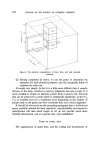
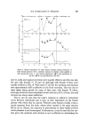
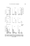
CHEMISTRY OF HUMAN HAIR CUTICLE--Ill 297 but insoluble material remaining after long term trypsin digestion was almost identical in composition to that remaining after 2 h digestion with pronase. (d) Endocuticle subfractions A1 and A2 From our study of the gravimetric course of digestion of whole cuticle with trypsin, two components dissolving at different rates were identified. Since we believe that long term trypsin treatment dissolves the effete cuticle nucleus, it follows that the two new fractions identified are subcomponents of this nucleus. That this is so is in agreement with the morphologic progression of trypsin diges- tion observed with the electron microscope. The component of the cuticle which dissolves in trypsin after 9 h and that which dissolves after treating with trypsin for a further 10 days have been designated endocuticle A1 and A2 respectively (i.e. ENDO A1 and A2). The amino acid analyses of these two subcomponents of the nucleus are shown in columns 5 and 6 of Table I. As would be expected the analyses of A1 and A2 are complementary with respect to the whole ENDO A fraction. In particular A1 contains significantly higher concentrations of aspartic acid, glutamic acid, leucine, tyrosine, phenylalanine and arginine and lower con- centrations of serine, proline, glycine, valine and cystine + cysteic acid than A2. It is also worth pointing out that the analysis for A1 is most extreme from that of whole cuticle. The nuclei of normal viable mammalian cells contain two major types of pro- tein, namely the histone proteins generally characterized by their high basic amino acid content (7), and the non-histone proteins which are relatively rich in acidic amino acids (8). It is difficult to imagine what changes occur to the corresponding proteins from the nuclei of the matrix cells of the hair follicle during the keratin- ization process and certainly their ultimate fate following loss of the nucleic acids of these cells is not known. Perhaps the two fractions A1 and A2 originate from the histone and non-histone proteins of the cuticle cell nucleus modified during the keratinization process. (e) Endocuticle fraction B (ENDO B) From the foregoing it is clear that there is a further fraction of the endocuticle which is not digested by long-term trypsin treatments but which does dissolve in pronase. This component comprises about 15•o by weight of whole cuticle and about 43•o by weight of the endocuticle. The amino acid analysis for this fraction, which we have designated ENDO B, is shown in column 7 of Table I. It is clear from comparison with the ENDO A fraction that ENDO B contains the bulk of the cystine present in the whole endocuticle and that the concentration of aspartic acid is lower.
Purchased for the exclusive use of nofirst nolast (unknown)
From: SCC Media Library & Resource Center (library.scconline.org)