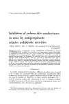
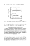
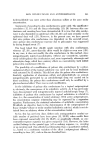
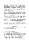
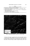
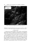
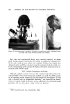
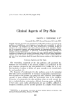
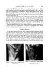
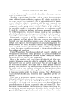
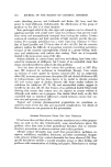
CLINICAL ASPECTS OF DRY SKIN 367 velocity. In 1962 Chernosky (2) found an identical clinical picture of dry skin in patients who were exposed to humid summertime weather and rela- tively dry refrigerated air conditioning. Clinical observations on dry skin fit with experimental studies concerning the nature of straum corneum hydra- ion, water binding, and water barrier properties, which have been thorough- ly reviewed by Idson (9). For over 50 years physicians have noted that soap and water (especially hot water) promote dryness of the skin, and they later noted that detergents and various solvents do the same thing (2, 4, 6, 10, 11). Numerous experi- mental studies have continned these clinical observations. Damage to water binding capacity is observed especially if the skin is exposed first to lipid solvents or detergents and then to water (12-14). Physical damage of the skin from clothing, pressure, rubbing, and scratch- ing have long been recognized as a causative in producing some of the clinical and histological changes in patients with dry skin. CLINICAL FEATURES In the early stages of dry skin, only a physicoche•nical disruption of the nonviable stratum corneum occurs. The earliest visible change, often not appreciable to the untrained observer, consists of a dull, greyish-white color with increased prominence of topographical lines that, upon dose inspection, look like an irregular mosaic (Fig. 3). Compare this change with normal appearing skin in the same anatomic area (Fig. 4). As changes in hydration of the corneum continue, the cohesiveness of the squames is partially lost, which allows a curling up of their edges and a network-like pattern of tiny -s, •¾:' ' ' %"::'•.:•:.':"a ß .. Figure 3. Earliest visible changes skin (see text) ß . •. ,:• •.. •.:::•-. $% :•.¾ •:i-:: .:.•./.. ..a.:.:. ..if 6....•. L•:.•.. ..• .•:• •:' .. ß •::,.. •.: .... '•"•!-•:•.,:.,,.,., .....: Figure 4. Normal skin of same anatomic location shown in Fig. 3

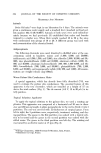
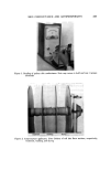
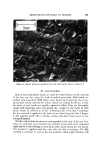
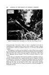
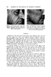
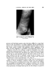
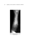
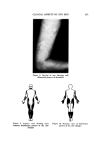
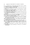
368 JOURNAL OF THE SOCIETY OF COSMETIC CHEMISTS Figure 5. Dry skin showing loosening of edges of surface squames and irregular pattern of chaps chaps (slits) in the skin surface (Fig. 5). Sometimes this change is more marked at t•ollicular orifices (Fig. 6). At first, the chaps involve only the stratum corneum, but they may progress deeper and become fissures that extend into the malpighian layer and even the dermis, thus causing biologic damage and stimulating an inflammatory response, which is clinically ob- served as erythema, edema, exudation, crusting, and occasionally, pur- pura (15). Loosening of entire squames from the surface results in shedding of dry scales. The skin surface looks and feels rough, and the patient may describe a feeling of loss of pliability when the skin is moved or stretched. Pruritus and a stinging sensation may be present, and various types of trauma from rubbing, scratching, and clothing friction aggravate these symp- toms and produce more surface damage, erythema, and edema, but rarely any vesiculation. A chronic lichenified dermatitis with pigmentary changes may ensue if the condition persists (Fig. 7). Secondary bacterial infection may also. occur. Distinct plaques of eczema may appear, sometimes with central clearing (Fig. 8), which resemble dermatophytosis. Some observers believe that trauma causes these patches to develop, but their nummular and avoid ap-
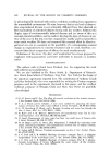
Purchased for the exclusive use of nofirst nolast (unknown)
From: SCC Media Library & Resource Center (library.scconline.org)