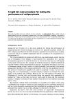
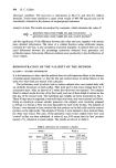
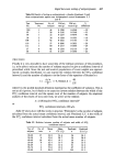
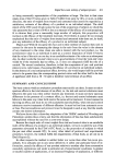
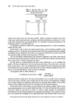
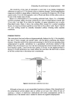
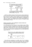
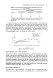
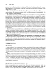
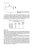
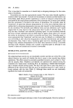
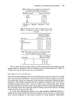
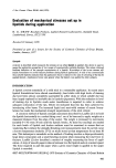
418 W. B. Davis and A.M. Rees-Jones In order to measure the rate of sweating, cylindrical cells (Fig. 3) are fixed to treated and control sites on the backs of subjects. 'Ambient' air (38.5øC 35•o RH) is drawn from the warm room and pumped into the cells at the rate of 150 ml per min through twelve 1'5 mm diameter holes situated concentrically inside the cells. The cells are held in dose contact with the skin by 3 cm wide strips of elastic thus preventing gross leakage. The outgoing air from each cell is directed through a humidity sensor*. The changes in capacitance of the sensor (related to humidity changes) are electronically processed and amplified before being recorded directlye' as percentage relative humidity. The air flow rate (150 ml/min) was found to be sufficiently fast to prevent the accumulation of liquid sweat on the test area. Figure 3. Hygrometric sweat measurement cell. The perspex cell directs air onto the sweating skin surface via a concentric ring of holes. The outgoing air has an elevated water content as a result of sweat evaporation. If any leakage occurs at the cell/skin boundary the air escaping will have picked up as much moisture as monitored outgoing air. Thus the product of the humidity rise and the ingoing flow rate are the relevant measurements. The outgoing flow rate is used to check for gross leakage. On entering the hot-room subjects were found to differ in their response to the thermal stress applied mainly in that the time required for sweating to increase to a consistent rate ranged from 10 to 30 min. The plateau values themselves varied from person to person and from day to day for each person. THERMOGRAPHIC MEASUREMENTS A sweat rate equal to 50 mg/min from the axillary vault draws 120 J/min from the skin surface to evaporate the sweat. When the room temperature is at or near skin temperature radiation causes no heat loss or gain, thus all the heat generated by the body should be lost by evaporation from the skin and lungs to maintain equilibrium. Under these con- ditions the skin surface is the coldest part of the environment. Localised cooling of the skin by evaporation contributes to thermal regulation, therefore it is reasonable to assume that there is less cooling of the skin over areas where an effective antiperspirant has been applied, i.e. the skin temperature is higher. Thermo- graphy, is in effect, the process of recording variations in intensity of long wavelength emissions from a surface in a mode of action similar to that of a visible wavelength television system. Hot areas emit more energy in the sensitive range of the instrument than cold areas so are thus displayed on a television screen as brighter areas in mono- chrome systems, or as a particular hue in colour systems displaying temperature vari- ations over the skin surface as a map (Figs. 4-7). * HP4 humidity & temperature probe, Lee Dickens Ltd, Kettering, England. •' Speedomax W Multipoint Recorder, Leeds & Northrup Ltd, Birmingham, England.

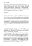
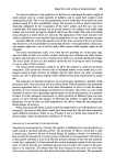
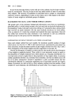
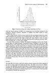
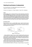
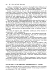
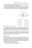
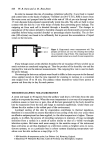
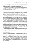
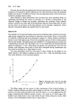
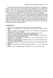
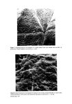
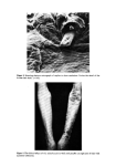
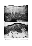
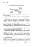
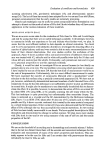
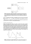
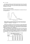
Figure 4. A thermogram of a non-sweating untreated back. A change of hue corresponds to 0'5 ø C. Figure 5. A thermogram of a thermally stressed back treated with antiperspirant. A cross of water (vertically) and product applied (horizontally) 20 cm below the shoulder is delineated by black square markers. There is a hot (green) strip of antiperspirant treated skin. 5 ø C scale length. Facing page 418
Purchased for the exclusive use of nofirst nolast (unknown)
From: SCC Media Library & Resource Center (library.scconline.org)