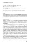
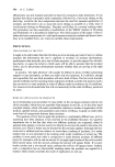
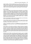
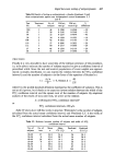
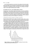
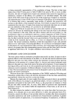
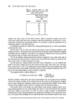
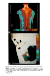
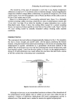
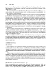
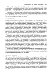
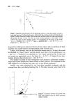
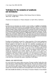
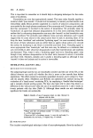
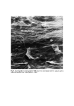
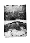
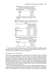
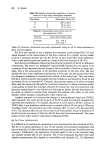
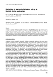
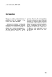
Figure 6. A thermogram of applied product evaporating in the axilla taken 10 min after application. The cool (coloured) cross is fading as the applications dry out. The surrounding areas appear white because they are hotter than the temperature range (1 ø C) covered by the ten colours. Figure 7. A thermogram of an axilla sweating during mental stimulation under ambient con- ditions. 5' C scale length. The normally hot central axillary region is 4 ø C cooler than the surrounding skin. Facing page 419

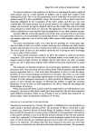
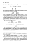
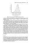
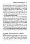
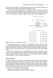
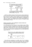
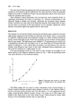
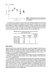
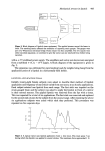
Evaluating the performance of antiperspirants 419 The sensitivity of this type of instrument is such that it can display temperature differences as small as 0.1 øC as distinct colour or intensity changes. Typical magnification ranges are available to image either a whole body or a portion of a finger on a 10 x 10 cm square screen. From the thermographic work of Parke and Reece (5) the axilla is seen to be one of the hottest areas of skin. Figure 4 is a thermogram of a non-sweating untreated back, Figure 5 is a thermally stressed (sweating) subject showing a relatively hot strip of antiperspirant treated skin 15 cm wide 3 cm high, 20 cm below his shoulder. (A green indentation into a cooler [blue-black] area). Figure 6 shows the cooling effect, caused for a period exceeding 10 min, by aqueous products applied in cross-form to the axillary vault Figure 7 shows 4øC local cooling caused by mentally stimulated axillary sweating under ambient conditions. PREDICTIONS The sweat gland, duct and orifice are diagrammatically displayed in Fig. 8. For simplicity the duct is drawn straight and parallel sided, with the gland comprising the bottom element. Using this model of the system and assuming it to be full but not operating when antiperspirant is applied, calculations by a geometrical sub-division method of the diffusive flux of aluminium ions down the duct indicated that several hundred ions could have reached a gland, 2 mm from the skin surface, in 20 min. As the applied product is drying (5-10 min) aluminium ions would diffuse into and down the duct from a source at the skin surface that is becoming more concentrated. Antiperspiran• ::::::,,: Skin ./.•,...•urføce .-• Duct Gland Figure 8. Geometrically subdivided sweat duct. Although we have not, as yet, accumulated conclusive evidence of the whereabouts of the antiperspirant activity (gland, duct or orifice) we do know that within 40 min of applying a solution of aluminium chlorhydrate (5/6 basic) antiperspirant activity is achieved even if the treated area is washed to remove the applied antiperspirant.
Purchased for the exclusive use of nofirst nolast (unknown)
From: SCC Media Library & Resource Center (library.scconline.org)