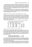
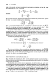
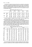
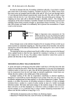
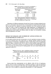
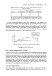
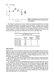
Evaluation of emollients and keratolytics 437 Table V. Results of use of bath oil to lower leg in six individuals on surfometer tracing area Time after exposure Area (cm2+ S.D.) in (min) tracing Before 8.5 + 2- 2 5 5.6+ 1.5* 60 8.2+ 1.9 240 6.9 + 2.3 * Significance difference fromcontrol 0-05 P0-02. Table VI. Results of use of HA on normal and dry skin subjects on surfometric tracing of SSB contrast with Table III Areas (cm2+ S.D.) in tracings from SSBs Time (min) Normal skin Dry skin Before 6.2+ 1.2 6.4+ 1.8 Immediately after 5.2 + 1.3 6.6 + 2.0 30 5.5+1-7 7.9+0.8 60 6.5+1.9 6.6+0.9 120 5.4+1.7 5.8+1.8 240 5.0+ 1.2 5.7+ 1.7 Table VII. Results obtained from SSBs after appli- cation of aqueous cream for 14 days Area (cm a + S.D.) in Time (days) n tracing from SSBs 0 8 12.9+3-7 7 9 12.0+1.8 14 8 11-7+2.0 Thus, it seems that the results of the use of HAs discernible at the surface, are also present within the substance of the SC although from the results presented in Tables V, VI and VII it may be that they are not as pronounced. (d) OTHER In Vivo TECHNIQUES There are two other techniques that we have used for the evaluation of HAs in vivo which have some functional significance. The first concerns the cohesive property of the SC. Our group have devised an instrument which measures intracorneal cohesion (coheso- graphy) (7). The method determines the force required to distract a disc of SC with a vertically orientated piston stuck to the skin surface with a cyanoacrylate glue. On the basis that 'hydration' was likely to alter the strength of the SC we used this instrument in the same study as that from which the results in Tables I and VII are derived, and these observations are set out in Table VIII. It is clear that although the results do not reach statistical significance there is a definite trend towards decreased intracorneal cohesion. We do not have as much experi- ence with this technique as with contour analysis for 'acute hydration' experiments.

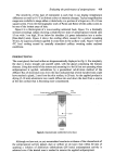
438 R. Marks Table VHI. Results obtained after application of aqueous cream for 14 days using cohesography technique Time (days) n Mean grams force + S.D. 0 7 109'1+49'0 7 8 94.4+34.3 14 5 85'2+ 19'9 Table IX. Cohesography results before and 3 h after application of HA Time (h) Grams force (Mean_+ S.D.) 0 (control) 222 + 69-2 3 157.7 + 64' 3 Table IJf, however, documents one such experiment using an oil in water emulsion in three normal subjects. We have also started to use a method that measures 'point penetrability' (8) and which depends on the measurement of the force required for a rapidly moving needle to move a measured distance into the SC. So far we have found that 'acute hydration' with a water soaked gauze pad results in a drop in the force required of 20-30•o. Both the above techniques utilise important physical properties of the SC as indicators of hydration. The next in vivo technique I want to briefly mention does not appear to be a measure of an important physical property, but is probably a function of many vari- ables. This is the measurement of the electrical properties of the skin. Clar, Her and Sturelle (9) have made significant contributions to this topic and this group claims that low frequency impedance is located almost entirely in the horny layer. They and others (10) have derived complex relationships between impedance and hydration based on the movement of ions in hydrated SC. Certainly, these groups appear to be able to evaluate HAs using this approach although the constraints necessary to obtain consistent results would appear to render the technique awkward for routine use. Our own experience with electrical measurements is very limited and although we believe that the techniques can be useful we intend to evaluate them alongside the methods with which we are more familiar, before making a commitment to their regular use. Another approach is to measure the changes in transepidermal water loss (TEWL) after hydration. The water barrier property of the SC is quite dramatically changed after relatively short hydration. For example, Quattrone et al. (1) report a 20-40• increase in TEWL after 5 min hydration which reverts to normal within a 30 min period. When an 'emollient cream' was used there was a 20• decrease after 1 h and a 10• decrease after 4-6 h. The decrease after the use of an occlusive HA presumably results in hydration of the SC from the movement of H•O into the SC from the living tissue beneath. This same author reviews other workers' experiences employing TEWL. (e) In Vitro TECHNIQUES It is difficult to be enthusiastic concerning in vitro techniques for the evaluation of HA. The SC is so utterly dependent in vitro on the ambient environmental temperature and relative humidity (11) that controlled environments and long periods of equilibration are necessary. Furthermore, the application of topical agents to the in vitro specimen does not accutely emulate the 'in use' situation. The in vitro techniques that have been used include measurements that depend on extensibility and elasticity (12, 13) differential
Purchased for the exclusive use of nofirst nolast (unknown)
From: SCC Media Library & Resource Center (library.scconline.org)