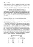
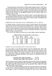
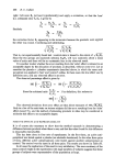
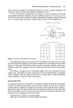
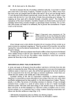
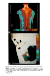
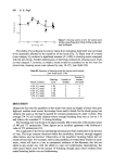
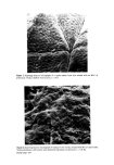
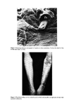
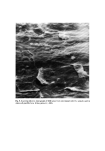
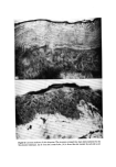
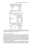
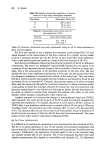
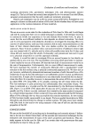
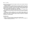
Evaluation of emollients and keratolytics 439 scanning calorimetry (14), gravimetric techniques (15), and photoacoustic spectro- scopy (16). The last of these has recently been adapted for in vivo use and I am told (Pines, personal communication) that the early results are extremely promising. Clearly such techniques may be useful to screen compounds before formulation or to attempt to dissect out the mode of action of HAs but I doubt whether they will have much application to the routine evaluation of these materials. DESCALING AGENTS (DAS) We are in an even worse state for the evaluation of DAs than for HAs and I could begin and end by saying that there are no useful techniques available. I will attempt, however, to summarise briefly our experience so far and indicate what further work, we plan. It seems that the most efficient method to date depends on clinical evaluation. Van Scott and Yu (17) used patients with ichthyotic disorders to investigate the descaling effect of a number of alpha-hydroxy acids and were certainly able to make recommendations on the basis of their clinical observations. Our own studies confirm the usefulness of this approach. Figure 4 shows a patient with a rare and severe form of ichthyosis whose right arm was treated with 6• salicylic acid in white soft paraffin twice daily for 10 days and whose left arm received just the vehicle. Fortunately, such patients are rare and it is just not a practical proposition to use this approach routinely. Clearly, it would be ideal to investigate DAs on normal human (or less ideally on animal) skin in vivo or in vitro. The real problem is knowing which parameter to measure. I have implied by my use of the term DA that the best kind of measurement would be on the rate of desquamation. Unfortunately, this is a most difficult measurement to make. We have examined the number of corneocytes liberated after a standardised 'scrub' stimulus to the skin surface using a specially constructed apparatus (18). We have not as yet, however, satisfactorily demonstrated increased numbers of cells liberated from a DA treated site. It may be that this technique is not sufficiently sensitive to pick up differences in normal skin. It might also be mandatory to use abnormally keratinised skin to demon- strate the effect. It is possible, however, to demonstrate the action of DAs on normal SC by either SEM (19) using SSBs, or by actually counting the cell strata within the SC. This last technique is quite promising but unfortunately does necessitate biopsy. The tissue is sectioned on a cryostat and the SC demonstrated by the McKenzie technique (20). Figure 5 is an SEM of SC taken after 10 days use of 6• salicylic acid in white soft paraffin and Fig. 6 shows cryostat sectioned skin treated by the same material compared to a vehicle treated specimen. Other workers (21) have also demonstrated this SC thinning effect of DAs although the way they accomplish this remains mysterious. In vitro testing of DAs has been totally unsuccessful in our hands. We have applied keratolytics to SSBs themselves and demonstrated no change in surface contour. We plan many more studies with DAs both in vitro and in vivo using measures of the rate of cell loss and the tensile properties of the SC. CONCLUSION We are on the threshold of an exciting era in dermatology and cosmetic science. I believe that in the not too distant future there will be accurate and convenient techniques for the evaluation and measurement of many of the skin's properties and functions. This cer- tainly appears to be the case for HAs and I am certain that with perseverance the same will shortly be true for DAs as well.

440 R. Marks REFERENCES 1 Quattrone, A. J. and Laden, K. physical techniques for assessing skin moisturization. J. Soc. Cosmet. Chem. 27 607 (1976). 2 Middleton, J. D. and Roberts, M. E. Efficacy of a skin cream containing pyrrolidone carboxylic acid in reducing the incidence of subclinical dry skin. In: Marks, R. and Dykes, P. J. The Ichthyoses. 177 (1978) MTP Press Ltd, Lancaster. 3 Garber, C. A. and Nightingale, C. T. Characterizing cosmetic effects and skin morphology by scanning electron microscopy. J. Soc. Cosmet. Chem. 27 509 (1976). 4 Bernstein, E. O. Scanning electron microscopy of skin topography before and after treatment. Ann. Meet. Soc. Cosmet. Chem. December 1974, New York City. 5 Marks, R and Dawber, R. P. R. Skin surface biopsy: an improved technique for the examination of the horny layer. Brit. •. Dermatol. 84 117 (1971). 6 Marks, R. and Pearse, A.D. Surfometry: a method of evaluating the internal structure of the stratum corneum. Brit. J. Derrnatol. 92, 651 (1975). 7 Marks, R., Nicholls, S. and Fitzgeorge, D. Measurement of intracorneal cohesion in man using in vivo techniques. J. Invest. Dermatol. 69 (3) 299 (1977). 8 Nicholls, S., King, C. S., Guibarra, E. and Marks, R. Measurement of point deformation (PD) of human skin in vivo: contribution of the stratum corneum. Ann. Meet. Eur. Soc. Dermatol. Res. Amsterdam (1978). 9 Clar, E. J., Her, C. P. and Sturelle, C. G. Skin impedance and moisturization. J. Soc. Cosmet. Chem. 26 337 (1975). 10 Edelberg, R. Relation of electrical properties of skin to structure and physiologic state. J. Invest. Dermatol. 69 324 (1977). 11 Agache, P., Boyer, J.P. and Laurent, R. Biomechanical properties and microscopic morphology of human stratum corneum incubated in a wet pad in vitro. Archiv. fiir Derrnatol. Forschung. 246 271 (1973). 12 Middleton, J. D. The effect of temperature on extensibility of isolated stratum corneum and its relation to skin chapping. Brit. J. Derrnatol. 81, 717 (1969). 13 Ferguson, J. and Agache, P. Influence of site, storage and trypsin treatment on the mechanical properties of the stratum corneum. J. Invest. Derrnatol. 68, 256 (1977). 14 Miller, D. L. and Wildanauer, R. H. Thermomechanical probes for the analysis of physical properties of stratum corneum. J. Invest Dermatol. 69 297 (1977). 15 Laden, K. & Spitzer, R. Identification of a natural moisturizing agent in skin. J. $oc. Cosmet. Chern. 18 351 (1967). 16 Rosencraig, A. and Pines, E. Stratum corneum studies with photoacoustic spectroscopy. J. Invest. Derrnatol. 69 296 (1977). 17 Van Scott, E. J. and Yu, R. J. Control of keratinization with alpha-hydroxy acids and related compounds. I. Topical treatment of ichthyotic disorders. Arch. Derrnatol. 110 586 (1974). 18 Nicholls, S. and Marks, R. Novel techniques for the estimation of intracorneal cohesion in vivo. Brit. J. Derrnatol. 96 595 (1977). 19 Davies, N. and Marks, R. Studies on the effect of salicylic acid on normal skin. Brit. J. Derrnatol. 95 187 (1976). 20 Mackenzie, I. C. and Linder, J. E. An examination of cellular organization within the stratum corneum by a silver staining method. J. Invest. Dermatol. 61 245 (1973). 21 Huber, C. and Christophers, E. 'Keratolytic' effect of salicylic acid. Arch. Derrnatol. Res. 257 293 (1977).
Purchased for the exclusive use of nofirst nolast (unknown)
From: SCC Media Library & Resource Center (library.scconline.org)