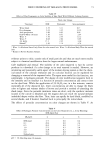
PROTEINS OF HAIR 99 (2) (3) (4) (5) (6) (7) (8) (9) (10) (11) (12) (13) (14) (15) (16) (17) (18) R. C. Marshall, Changes in wool low sulfur and high sulfur protein components following chemical defleecing, Text. ResearchJ., 51, 384-388 (1981). R. D. B. Fraser, T. P. MacRae, and G. E. Rogers, Keratins, Their Composition, Structure and Biosyn- thesis (Thomas, Springfield MA, 1972), pp. 30-55. P.M. Steinert and G. E. Rogers, Characterization of the proteins of guinea pig hair and hair follicle tissue. Biochem. J., 135, 759-771 (1973). R. C. Marshall, Genetic variation in the proteins of human nail, J. Invest. Dermatol,, 75, 264-269 (1980). R. D. B. Fraser and T. P. MacRae, Current Views on the Keratin Complex From the Skin of Vertebrates, R. I. C. Spearman and P. A. Riley, Eds. (Acad. Press, 1980), pp. 67-86. J. M. Gillespie, The dietary regulated biosynthesis of high sulfur wool proteins, Biochemo J., 98, 669-677 (1966). L.J. Wolfram, "The Reactivity of Human Hair. A Review," in Hair Research, Status and Future Aspects, C. E. Orfanos, W. Montagna, and G. Stiittgen, Eds., 1979, pp. 479-500. J. M. Gillespie and R. C. Marshall, A comparison of the proteins of normal and trichothiodystrophic human hair, J. Invest. Dermatol., 80, 195-202 (.1983). H. P. Baden, L. D. Lee, and J. Kubilus, A genetic electrophoretic variant of human hair--polypep- tides, Am. J. Hum. Genet., 27, 472-477 (1975). U. K. Laemmli, Cleavage of structural proteins during the assembly of the head of bacteriophage T4, Nature, 227, 680-685 (1970). B. J. Davis, Disc electrophoresis II. Method and application to human serum proteins, Ann. N.Y. Acad. Sci., 121, 404-427 (1964). R. A. Laskey and A.D. Mills, Quantitative film detection of 3H and •4C in polyacrylamide gels by fluorography, Eur. J. of Biochem,, 56, 335-341 (1975). M. M. Bradford, A rapid and sensitive method for the quantification of microgram quantities of protein utilizing the principle of protein-dye binding, Anal. Biochem., 72, 248-254 (1976). J. P. Ortonne and J. Thivolet, "Hair Melanin and Hair Color," in Hair Research, C. F. Orfanos, W. Montagna, and G. St/ittgen, Eds. (Springer-Verlag, Berlin, Heidelberg, 1981), pp. 146-162. A. Rook, The clinical importance of "weathering" in human hair, Br. J. Derm., 95, 111 (1976). C. R. Robbins and M. K. Bahl, Analysis of hair by electron spectroscopy for chemical analysis. J. Soc. Cosmet, Chem., 35, 379-390 (1984). W. G. Crewther, R. D. B. Fraser, F. G. Lennox, and H. Lindley, The chemistry of keratins, Advan, Protein Chem., 20, 191 (1965).

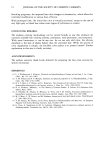
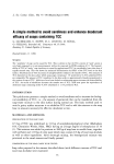
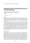
j. Soc. Cosmet. Chem., 40, 101-107 (March/April 1989) The role of antioxidants in skin immune reactions: The use of flow cytometry to determine alterations in la-positive epidermal cells in allergic contact dermatitis LAWRENCE A. RHEINS,* RICHARD A. MORAVEC, MICHAEL L. NORDLUND, LINDA S. TRINKLE, and JAMES J. NORDLUND, Department of Dermatology, University of Cincinnati College of Medicine, Cincinnati, OH 45267. Synopsis Earlier experiments from our laboratory revealed that common parasubstituted phenolic compounds such as monobenzyl ether of hydroquinone (MBEH), butylated hydroxytoluene (BHT), and butylated hydroxyani- sole (BHA), ubiquitous compounds found in medications, cosmetics, foods, and various industrial products, can alter Ia+ Langerhans cells (epidermal macrophages) and epidermal immune responsiveness (i.e., exacerbate allergic contact dermatitis) when topically applied to the epidermis of various strains of mice. The current experiments demonstrated that a precise and sensitive instrumental approach, fluorescent activated cell sorting (FACS), could be used to quantify changes more precisely in the expression of the membrane-bound Ia antigen found on epidermal antigen-presenting-Langerhans cells during contact hyper- sensitivity reactions. Further, this technique can be used to screen potential vehicle and/or medicament allergens as likely sources for allergic contact dermatitis at home and in the work place. INTRODUCTION Earlier studies from our laboratory have documented that topical applications of common antioxidants such as monobenzyl ether of hydroquinone (MBEH), butylated hydroxyanisole (BHA), and butylated hydroxytoluene (BHT), ubiquitous compounds found in medications, cosmetics, food, and industrial rubber products, can alter the contact hypersensitivity responsiveness (CHS) (e.g., allergic contact dermatitis, ACD) in various mice strains when epicutaneously applied (1). These compounds, when topi- cally applied for a short period of time (i.e., daily for five days) to various epidermal sites, can alter the density (cells/ram 2) of Ia + (immune associated antigen)/ATPase + Langerhans cells (LC) (epidermal immune macrophages). Further, this increase or de- crease in identifiable Langerhans cells could be correlated with alterations in CHS re- sponsiveness following sensitization and challenge with common allergens/haptens (i.e., dinitrofiuorobenzene, oxazalone) (1). Other studies suggest that the mechanism responsible for the alteration of Ia + Langerhans cell density and functional CHS reactivity following topical antioxidant treatment may be mediated via arachidonic acid (AA) and/or its metabo- lites (i.e., prostaglandins, thromboxanes, leukotrienes)(1,2). * Reprint requests should be sent to: Lawrence A. Rheins Ph.D., Procter & Gamble Co., Ivorydale Tech- nical Center, Cincinnati, Ohio 45217 101
Purchased for the exclusive use of nofirst nolast (unknown)
From: SCC Media Library & Resource Center (library.scconline.org)