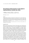
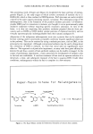
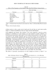
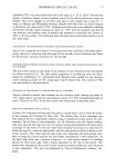
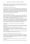
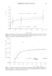
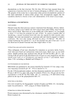
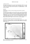
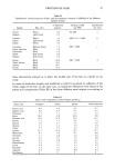
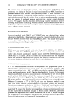
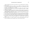
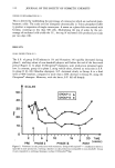
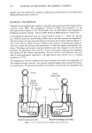
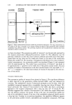
120 JOURNAL OF THE SOCIETY OF COSMETIC CHEMISTS profiles were then analyzed by a dynamic mathematical model based on the bilayer skin diffusion/bioconversion model (5). MATERIALS AND METHODS Vitamin E (o•-tocopherol) and vitamin C (ascorbic acid) were provided by Sigma Chem- icals (St. Louis, MO). The radiolabeled vitamin E ((3H)2-3,4o•-tocopherol, 10-40 mCi/mmol) and vitamin C L-[1-•4C]-ascorbic acid, 10 mCi/mmol) were supplied by Hoffmann-La Roche (Nutley, N J) and NEN Research (Massachusetts), respectively. A full-thickness abdominal skin of a female hairless mouse (5-7 weeks old, Jackson Lab. HRS/J strain) was excised freshly before the in vitro skin penetration experiment. The skin sample was then mounted between the donor and receptor halfcells (Figure 1). The in vitro system, which has been calibrated with respect to hydrodynamic character- istics (6), assures the intrinsic skin penetration (7) under the present experimental con- dition. The donor and receptor solutions (Table I) were then charged in each cell com- partment. At appropriate time intervals, 30 }xl of the receptor solution was withdrawn and assayed for the vitamin concentration. Six sets of the in vitro diffusion cells were used in each penetration experiment. All experiments were carried out at a constant temperature (37øC). The degradation of both vitamins in the donor solution was found to be negligible. In the receptor solution, however, the vitamins degraded appreciably during the penetra- tion experiment. The penetration profile was therefore corrected for the degradation by ( ,••Stopper • St•rnng [• j (10 mm dia.,
Platform 4 mm hgt ) .., '•"-• / Slar-hne•d 35ml .... capacity Filling & Sampling Port Skin lIGHT SEAL DONOR (Left) HALF-CELL RECEPTOR (Righi) HALF-CELL Figure 1. In vitro skin penetration apparatus used in this study.

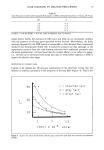
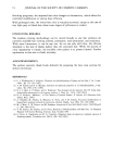
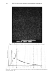
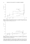
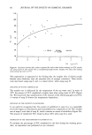
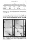
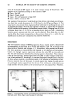
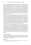
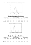
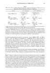
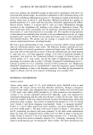
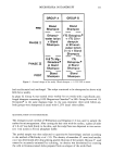
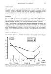
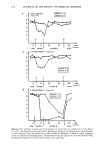
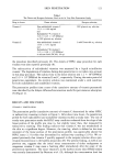
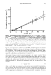
SKIN PENETRATION 121 Table I The Donor and Receptor Solutions Used in the In Vitro Skin Penetration Study Drug in donor Donor solution Receptor solution Vitamin C Non-radiolabeled vitamin C 50% glycerin aq. solution 12.11 -+ 0.86 mg/ml and Radiolabeled •4C-vitamin C 5.93 --- 0.25 X 10⁵ DPM/ml in 50% glycerin aq. solution. Non-radiolabeled vitamin E 13.81 -+ 0.51mg/ml and Radiolabeled 3H-vitamin E 1.23 + 0.09 X 10⁵ DPM/ml in silicone fluid (DC360, 20 cp) Vitamin E 5 mM Tween-80 aq. solution the procedure described previously (8). The details of HPLC assay procedure for each vitamin were also reported previously (8). The radioactivity of radiolabeled vitamins was measured by a liquid scintillation counter. The degradation of vitamins during skin penetration is not taken into account in this assay procedure. The radioactivity in the donor solution was 1.2 X 10 5 DPM/ml and 5.9 X 10 5 DPM/ml for vitamins E and C, respectively. During the entire period of penetration experiment, the receptor solution was maintained under a sink condition for both radiolabeled and nonlabeled vitamins. The penetration profiles (time course of the cumulative amount of vitamin penetrated) were described by the bilayer diffusion/bioconversion model for percutaneous absorption (5) (Figure 2). RESULTS AND DISCUSSION VITAMIN C PENETRATION The penetration profile (cumulative amount) of vitamin C determined by either HPLC or radioactivity counting is shown in Figure 3. After about nine hours, the penetration profile for both radiolabeled and nonlabeled vitamins reached a steady state. The rate of steady-state penetration under the HPLC assay condition evaluated from the slope of the linear portion of the profile was close to, but slightly lower than, that measured by radioactivity counting. This finding indicates that vitamin C was not metabolized in the skin to a significant degree. However, the time-lag, which is defined as the time intercept of the linear portion of the penetration profile, was appreciably shorter for HPLC assay (1.3 h) than that determined by radioactivity counting (3.9 h). We re- ported previously that vitamin C quickly appeared in the receptor solution after a pro- vitamin bioconversion in the hairless mouse skin (8). The present finding, as well as our previous one, suggests that the initial tissue concentration of either endogenous or exogenous vitamin C may cause a bursting effect immediately after the onset of the penetration experiment. The tissue vitamin C in the hairless mouse skin (HRS/J strain) was recently demonstrated by Buettner et al. (9), although the concentration level has
Purchased for the exclusive use of nofirst nolast (unknown)
From: SCC Media Library & Resource Center (library.scconline.org)