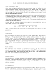
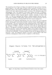
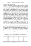
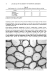
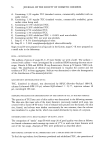
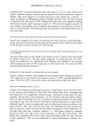
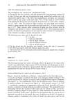
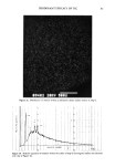
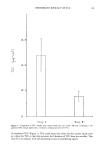
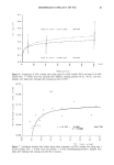
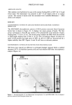
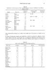
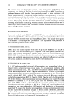
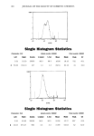
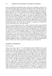
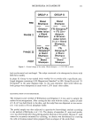
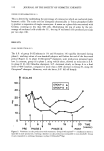
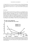
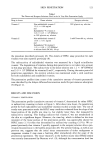
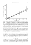
ANTIOXIDANTS IN IMMUNITY 105 Table I Effect of AA, BHA, and BHT on Mean Phenotypic Ia q- Epidermal Cell Expression: Effects of Low- and High-Dose AA With Antioxidant on Mean Ia + Expression Control % Ia+ Expression % Ia + Expression Diluent (DMSO)* 3.81 Arachidonic acid Arachidonic acid 66 ng AA 4.72 666 AA ng 1.42 (+ 24%) (- 63%) BHA AA + BHA AA + BHA BHA diluent (DMSO) 66 ng AA + 5 •xg/ml 666 ng AA + 5 •xg/ml + BHA 5 •xg/ml 2.88 BHA 3.62 BHA 2.11 (q' 25%) (-27%) BHT BHT + AA BHT + AA BHT diluent (DMSO) 66 ng AA + 2.5 I-tg/ml 666 ng AA + 2.5 •xg/ml + BHA 2.5 •xg/ml 3.99 BHT 4.44 BHT 3.00 (q. 11%) (-25%) * DMSO: 100 Ixl was used as delivery for BHT, BHA, and AA concentrations. 4 ( 105 unenriched epidermal cells were plated into 24-well polystyrene plates containing antioxidant- supplemented RPMI-1640 followed by a pulse of either low- or high-dose AA. Following a 48-hour incubation at 37øC, 5% CO2, the cells were harvested and stained with appropriate primary and secondary fluorochrome-conjugated antibodies and then subjected to FACS analysis. There is an apparent biphasic dose effect on the mean expression of the Ia + antigen, suggesting that the dose/concentration of the antioxidant can contribute significantly to altering immune phenotypic marker expression. of common parasubstituted phenolic antioxidants, namely MBEH, BHT, and BHA, appear to alter the density of identifiable Ia+ Langerhans cells in various strains of mice. Further, these alterations in Ia+ Langerhans cells were dose-related and corre- lated with either enhancement or suppression of the contact hypersensitivity response in these mice strains (1). In addition, this effect could be totally reproduced by using a biologically relevant molecule, namely arachidonic acid. However, these earlier in vivo experiments performed in our laboratory were "gross," using the imprecise mouse ear swelling assay, which lacks the precision and sensitivity needed to validate our earlier functional CHS observations. The current studies were carried out using a more precise methodology, that of fluores- cent-activated cell sorting, to determine whether it could serve as a screening technique to determine which of these ubiquitous antioxidants/preservatives may contribute to Figure 1. Changes in Langerhans cell density and Ia expression following topical treatment with MBEH: Increase in density (number) and class II MHC product (Ia antigen) of unenriched murine Langerhans cells following five days of in vivo topical treatment with diluent (a) or MBEH (b). To identify Ia q. Langerhans cells, dual labeling immunofluorescence was performed using fluorescein isothiocyanate (FITC)-labeled anti-Fc receptor and monoclonal anti-class II MHC streptavidin phycoerythrin red (PE) antibodies. In other words, two cell membrane markers were used to identify Langerhans cells from keratinocytes (i.e., the Ia marker and the Fc marker are found only on Langerhans cells in normal epidermis). The percentage of identifiable Iaq, Langerhans cells increased significantly from 1.4% to 3.3% in fluorescent intensity as shown in the histogram, Figures la and lb, respectively. Arrows indicate positive signal gated regions (i.e., FACS is able to detect fluorochrome marker expression). The mean fluorescent intensity (i.e., mea- surement/density of Ia marker) also increased from 69.51% (diluent) to 72.99% (treated). X-axis denotes fluorescent intensity y-axis denotes the number of events.

106 JOURNAL OF THE SOCIETY OF COSMETIC CHEMISTS exacerbated allergic contact dermatitis via the alteration of Ia+ epidermal immune cells. In the present study we were able to demonstrate that a short-term application of MBEH could significantly increase the density of Langerhans cells (1.4% to 3.3%) as well as the mean phenotypic expression of the Ia antigenic marker. Several investigators have reported that the increase in Ia q- antigen or analogue in man, HLA-DR, can be directly correlated with an intensified allergic contact dermatitis reaction. Further, our current studies examine more ubiquitous antioxidants, namely BHT and BHA, using an in vitro cell culture system. Results from these experiments strengthen our earlier findings that antioxidants can play a role in altering epidermal immune reactivity. Further, this reponse could be reproduced using a biologically relevant molecule, ara- chidonic acid. In summary, we have presented the results of experiments which demonstrate the use of FACS as a sensitive and precise tool to measure small yet significant identifiable changes in epidermal immune molecules residing in the epidermis. This technique, along with the use of human epidermal cell suspensions obtained from various sources (i.e., sur- gical specimens), theoretically provides one with a technique to determine which cos- metics and/or pharmaceutical formulations may alter the epidermal immune homeo- stasis, thereby potentially leading to exacerbated allergic contact dermatitis both in the home and in the workplace. ACKNOWLEDGMENTS This study was supported in part by NIH Grant AM25252, NIOSH Grant ROI OH02091, and the Elma Margaret Lapp Foundation, Cincinnati, Ohio. The authors are grateful to Ms. Catherine Goldschmidt for her assistance in the prepara- tion of the manuscript. REFERENCES (1) L. A. Rheins and J. j. Nordlund, Modulation of the population density of identifiable epidermal Langerhans cells associated with enhancement or suppression of cutaneous immune reactivity. J. Immunol., 136, 867-876 (1986). (2) L. A. Rheins, E. M. Young, M. L. Nordlund, R. B. Berning, and J. J. Nordlund, Rapid induction of Thy-1 antigenic markers on keratinocytes and epidermal immune cells in the skin of mice fol- lowing topical treatment with common preservatives used in topical medications and in foods, J. Invest. Dermatol., 87, 489-494 (1987). (3) C. A. Janeway, A. K. Bottomly, J. Babich, P. Conrad, S. Conzen, et al., Quantitative variation in Ia antigen expression plays a central role in immune regulation, Immunol. Today, 5, 99-104 (1984). (4) L. K. Roberts, G. G. Krueger, and R. A. Daynes, Correlation between the inducible keratinocyte expression of Ia and the movement of Langerhans cells into the epidermis, J. Immunol., 134, 3781-3784 (1985). (5) L. K. Roberts, G. J. Spangrude, R. A. Daynes, and G. G. Krueger, Correlation between keratino- cyte expression of Ia and the intensity and duration of contact hypersensitivity responses in mice, J. Immunol., 135, 2929-2936 (1985). (6) V. B. Morhenn, B. J. Nickoloff, and J. N. Mansbridge, Induction of the synthesis of Triton-soluble proteins in human keratinocytes by gamma interferon, J. Invest. Dermatol., 85, 27-29 (1985). (7) L. A. Rheins, L. Barnes, S. Amornsirpanitch, C. E. Collins, and J. J. Nordlund, Suppression of the
Purchased for the exclusive use of nofirst nolast (unknown)
From: SCC Media Library & Resource Center (library.scconline.org)