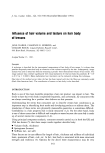
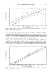
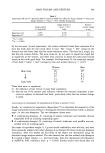
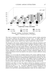
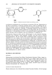
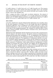
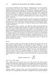
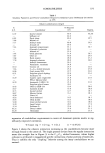
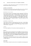
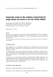
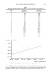
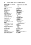
362 JOURNAL OF THE SOCIETY OF COSMETIC CHEMISTS HOCH 0 ¸ OH H H I HO H OH 0 CHOH (a) (b) Figure 1. Chemical structures of arbutin (a) and kojic acid (b). or melanocyte proliferation due to such factors as ultraviolet (UV) rays. Pigmentation in the skin is prevented by 1) reducing tyrosinase activity, either by inhibiting the syn- thesis of tyrosinase, which is an important enzyme for melanin synthesis, or by using an antagonist of the substrate for tyrosinase 2) decreasing melanocyte functions such as proliferation or decreasing melanin production by using agents that are cytotoxic to melanocytes 3) reducing dopa to prevent its auto-oxidation and 4) suppressing inflam- matory reactions, such as erythema, that occur following UV irradiation (3). Hydroquinone drugs are widely used by dermatologists to treat abnormal hyperpig- mentation such as freckles, senile lentigines, melasma, and other forms of melanin hyperpigmentation. The use of these drugs in bleaching creams is permitted in some countries (4,5), but in Japan, their use as cosmetic components is prohibited, as they have been reported to cause irritation and dermatitis under certain conditions (6,7). The selective depigmenting effect of hydroquinone on melanocytes, in association with its chemical structure, has been studied by biochemical methods and electron microscopy (8-12). Its effects are considered to be due to the inhibition of tyrosinase in melanocytes (8,9) and to its cytotoxicity to the melanocytes (10-12). Depigmentation and its mech- anism vary among arbutin, kojic acid (1), ascorbic acid (1), and finoleic acid. To compare the depigmenting effects of these agents, we evaluated their effects on tyros- inase activity, cell viability, and melanin synthesis in cultured human melanocytes, using hydroquinone as a positive control. MATERIALS AND METHODS MATERIALS Hydroquinone was obtained from Mitsui Petrochemical Industries (Tokyo, Japan) ar- butin was obtained from Nippon Fine Chemical Co. Ltd. (Osaka, Japan) kojic acid was purchased from Sigma Chemical Co. (St. Louis, MO) ascorbic acid and linoleic acid were purchased from Wako Junyaku Co. (Osaka, Japan). MCDB153 medium was purchased from Sanko Junyaku Co. Ltd. (Tokyo, Japan). And MTT assay kit was from Chemicon International, Inc. (Temecula, CA). CULTURE OF HUMAN MELANOCYTES Human melanocytes were obtained from neonatal Caucasian foreskins. Seventh passage

IN VITRO EFFECT OF WHITENING COSMETICS 363 melanocytes were cultured for about two weeks in MCDB153 medium containing 0.13 mM CaCI 2, 10 ng/ml epidermal growth factor (EGF), 5 •g/ml insulin, 0.5 •g/ml hydrocortisone, 1 ng/ml recombinant basic fibroblast growth factor (rbFGF), 10 ng/ml phorbol 12-myristate, 13-acetate (PMA) and 0.2% v/v of bovine pituitary extract. After replacement of the medium with MCDB 153 without rbFGF and PMA, the cells were cultured for a further two days. The cultured melanocytes were placed in 96-well plates at a density of 12,500 cells/cm 2. Two plates, one for the measurement of tyrosinase activity and the other for the measurement of cell viability, were cultured for one day. Hydroquinone and linoleic acid at final concentrations of 0.001, 0.005, 0.01, and 0.05 mM, and arbutin, kojic acid, and ascorbic acid at final concentrations of 0.01, 0.05, 0.10, 0.50, 1.0, and 5.0 mM, were added to triplicate wells and cultured at 37øC for three days. CELL VIABILITY ASSAY The cell viability in the presence of each agent was evaluated by the MTT test (13). After culture, phosphate buffer (pH 7.4) containing 5 mg/ml MTT (3-(4,5-dimethylthiazol- 2-yl)-2,5-diphenyl tetrasolium bromide) was added to each well. The plate was incu- bated at 37øC for 4 hours, after which isopropanol containing 0.04 N HC1 was added. Viable cells formed dark blue formazan by cleaving MTT with mitochondria. After 30 minutes, absorbance was measured at 570 nm using 655 nm as a reference. To eliminate agent interference in the measurement, wells containing each test agent alone were incubated and reacted with MTT. Cell survival was calculated from the absorbance. TYROSINASE ACTIVITY ASSAY The cells were washed with PBS and lysed with 45 •1 of 1% Triton-X/PBS. After vibration, 5 •1 of 10 mM L-DOPA was added to the wells. After incubation of the plates at 37øC for 60 min, absorbance was measured at 475 nm in a Model-3550 ELISA Reader (Bio-Rad Lab., Richmond, CA). The absorbance values were compared with a standard curve obtained with mushroom tyrosinase (Sigma Chemical Co., St. Louis, MO) the standard curve was linear within the range of experimental values. The coefficient of correlation was determined as 0.999. MELANIN ASSAY Melanin content was determined according to the method described by Oikawa (14), which we modified. Briefly, human melanocytes (40,000 cells/well) cultured in 12-well plates in 1.0 ml of medium were cultured with arbutin (0.5 mM), kojic acid (0.5 mM), and ascorbic acid (0.5 mM) at 37øC for three days. The culture solution was aspirated, and 1.0 ml of 2.5% trypsin solution was added to each well. The cells were detached, placed in an Eppendorf tube, and centrifuged at 1,000 rpm in a Hitachi CR 15 refrigerated centrifuge to obtain cell pellets. The pellets were mixed with 5% trichlo- roacetic acid, agitated well, and centrifuged at 10,000 rpm to deposit melanin. The melanin sediment was washed with PBS and mixed with 1N NaOH for dissolution, and the absorbance at 475 nm was measured. The amount of melanin per cell was calculated
Purchased for the exclusive use of nofirst nolast (unknown)
From: SCC Media Library & Resource Center (library.scconline.org)