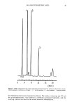
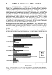
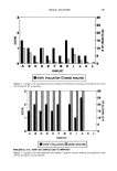
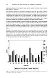
j. Soc. Cosmet. Chem., 43, 85-92 (March/April 1992) Protective effect of a topically applied anti-oxidant plus an anti-inflammatory agent against ultraviolet radiation-induced chronic skin damage in the hairless mouse D. L. BISSETT, R. CHATTERJEE, and D. P. HANNON, The Procter & Gamble Company, Miami Valley Laboratories, Cincinnati, OH 45239-8707. Accepted January 24, 1992. Synopsis Female albino hairless mice (Skh:HR-1) exposed chronically to sub-erythemal doses of ultraviolet radiation develop visible skin changes, histological alterations, and tumors. Topical treatment of mice with binary combinations of an anti-oxidant (alpha-tocopherol, ascorbic acid, or 2,4-hexadien-l-ol) and an anti- inflammatory agent (hydrocortisone, naproxen, or ibuprofen) prior to each UVB radiation exposure reduced significantly the severity of the observed photodamage events. The combinations provided protection additive of the effects of the individual components. UVA radiation-induced photodamage was inhibited effectively by the anti-inflammatory agent alone. Addition of an anti-oxidant did not increase this level of protection. INTRODUCTION Activated oxygen species and oxygen radicals have been implicated in ultraviolet (UV) radiation damage to skin (1-4). These species, in particular superoxide and singlet oxygen, are probably involved in chronic photodamage, since topical anti-oxidants that scavenge these species are photoprotective in the hairless mouse (2,3). Also, oxygen radicals are probably a primary factor in chronic photodamage, since chelators, which prevent iron-catalyzed production of oxygen radicals, are dramatically photoprotective in the mouse (4). Inflammatory cells also appear to play a role in photodamage. With chronic UV radi- ation exposure of mouse skin, there is an increase in dermal cellularity, including inflammatory cells (5,6). We have observed that topical anti-inflammatory agents are protective against chronic photodamage (7). This suggests that the inflammatory cell infiltrate contributes to the damage, although it is not clear what specific role these cells play in the damage process. 85

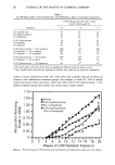
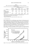
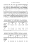
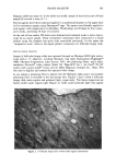
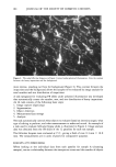
86 JOURNAL OF THE SOCIETY OF COSMETIC CHEMISTS Since either anti-oxidants or anti-inflammatory agents alone provide protection, it is likely that a combination of the two would have greater effectiveness. In this report, we describe the protective effect in hairless mice of the combination against UV radiation- induced chronic skin damage. The degree of protection is quantified using both histo- logical and visible perception methods. MATERIALS AND METHODS ANIMALS Female albino hairless Skh:HR-1 mice were obtained from the Temple University Health Sciences Center, Philadelphia, PA. They were housed five to a cage in a room with controlled temperature and humidity and with a 12-hour light/darkness cycle. They were given a standard diet and water ad libitum. Mice were approximately 10 weeks old at the start of experimental work. At the end of experimental work, they were sacrificed by CO 2 asphyxiation. IRRADIATION AND TOPICAL TREATMENT The procedure for irradiation of the dorsal skin of mice with UVB or UVA radiation has been described previously (6,8). Briefly, mice were irradiated individually under a bank of four Westinghouse FS-40 suniamps (UVB radiation, peak of emission near 315 nm) or four General Electric F-40 black lights (UVA radiation, no detectable emission below 340 nm). The output of the sources was monitored with an International Light (New- buryport, MA) model IL1350 radiometer with model SED240 (UVB) and SED015 (UVA) sensors. Mice were irradiated three times weekly (Monday, Wednesday, and Friday) with 30 mJ/cm 2 UVB radiation per exposure (approximately one half the mouse MED) or five times weekly (Monday-Friday) with 15 J/cm 2 UVA radiation per expo- sure. For mice receiving topical treatment with anti-inflammatory agents and/or anti- oxidants, the dorsal skin of the mice (n = 10 per group neck to tail area) was treated with 0.1 ml of test material solution two hours prior to each UV radiation exposure. Solutions were w/v% (pH 6-7) in an ethanol:propylene glycol:water vehicle [typically 2' 1' 1 (v:v:v)], the ratio of the components varying somewhat, depending upon the solubility of the particular test material. The test solution was delivered to the skin using a Pipetman © (Rainin Instrument Co., Woburn, MA) and spread evenly over the dorsal skin with the flat edge of the disposable piper tip. VISIBLE SKIN GRADING Skin wrinkling (UVB radiation-induced event) and skin sagging (UVA radiation- induced event) in hairless mice were assessed as described previously (6-8). Briefly, wrinkling and sagging were evaluated using grading scales (0-3, with half-grade in- crements), where 0 is normal and 3 is the maximum visible skin change observed in our work (6). Skin lesions were diagnosed as tumors if they were circular, red, raised, and greater than 1 mm in diameter. These lesions were counted.
Purchased for the exclusive use of nofirst nolast (unknown)
From: SCC Media Library & Resource Center (library.scconline.org)