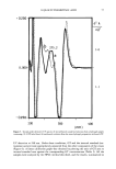
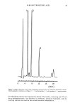
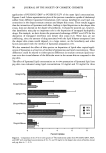
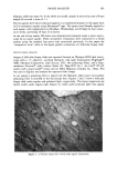
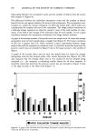
J. Soc. Cosmet. Chem., 43, 93-100 (March/April 1992) Influence of formulation factors on the deposition of liposornal components into the different strata of the skin J. DU PLESSIS, K. EGBARIA, and N. WEINER, College of Pharmacy, University of Michigan, Ann Arbor, MI 48109-1065. Received August 25, 1991. Synopsis The effects of lamellarity and total lipid concentration on the deposition of liposomal components into the various skin strata were determined using in vitro diffusion experiments. Large unilamellar vesicles, mul- tilamellar vesicles, and dehydration/rehydration vesicles composed of egg lecithin, cholesterol, and cho- lesteryl sulfate at lipid concentrations of 10, 25, and 50 mg/ml were tested. The results suggest that mixing and interaction of liposomal bilayers with the stratum corneum is extensive and that the skin is capable of incorporating large amounts of lipids when they are applied in a bilayer configuration. For each type of liposome tested, the amount of lipid deposited in the deeper skin strata of pig skin was at least ten times higher than that deposited in mouse skin. Lamellarity had little effect on the deposition of liposomal components into the skin strata when the formulations were compared at equal lipid concentrations. INTRODUCTION Liposomes are microscopic vesicles composed of one or more lipid bilayers arranged in concentric fashion, enclosing an equal number of aqueous compartments (1). Various amphipathic molecules have been used to form the liposomes, and the method of preparation can be tailored to control their size and morphology. The classification of liposomes is often confusing and can be based on whether they contain only one (uni- lameliar) or more (multilamellar) bilayers, their size, or their method of preparation. Recently, a great deal of interest in the use of liposomes in skin gels or skin creams has been generated in the field of cosmetics. Phospholipids are widely used for topical applications in cosmetics and dermatology, since they have a high content of esterified essential fatty acids, the proper blend of which is believed to increase the barrier function of the skin and decrease water loss within a short period of time after application (2,3). The key ingredient that keeps the human skin soft and flexible is water. Skin, partic- ularly the horny layer, performs a significant protective role by providing, in addition to mechanical protection, a barrier against extraneous substances. This function of the horny layer is dependent on its elasticity, determined by the content of fats and inor- ganic salts, as well as by the hydration state. Increasing the skin humidity leads to an increase in its elasticity (5). 93

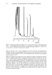
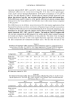
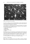
94 JOURNAL OF THE SOCIETY OF COSMETIC CHEMISTS Skin humidity is regulated to a large extent by lipids in the skin's horny layer. This complex lipid mixture is oriented, at least in part, as bimolecular leaflets (4). Liposomes have been employed in cosmetics and skin care products for several years with great success. However, few reports on the deposition of liposomal lipids into skin after topical application have been published (5-7). The objectives of the present study were to understand the nature of interaction of liposomal components with the skin by investigating the effect of liposome type and lipid concentration on deposition into the various strata of hairless mouse and pig skin using in vitro diffusion studies. MATERIALS AND METHODS Cholesterol (CH), cholesteryl sulfate (CS), and HEPES free acid were obtained from Sigma (St. Louis, MO). Egg lecithin (PC) was obtained from Avanti Polar Lipids (Birmingham, AL). ot-Tocopheral ((x-T) was obtained from Eastman Kodak (Rochester, NY). •4[C]-CS and 3[H]-CH were obtained from Amersham (UK). All other chemicals were of analytical grade. PREPARATION OF LIPOSOMES Multilamellar liposomes (MLV) containing PC:CH:CS at a molar ratio of 1:0.5:0.1 were prepared using the conventional film method (1). Briefly, the lipid mixtures-were dissolved in a 2:1 (v/v) mixture of chloroform and methanol. Trace amounts of 3[H]-CH and •4[C]-CS were incorporated in the phospholipid-based liposomes. The lipids and markers were deposited as a thin film in a round-bottomed flask by rotary evaporation under nitrogen. The flask containing the lipid film was stored in vacuum overnight to facilitate removal of residual solvents. The films were hydrated by the addition of an isotonic 0.05 M HEPES buffer, pH 7.4, with mild agitation at 45øC. The final concentrations of lipid were 50 mg/ml, 25 mg/ml, and 10 mg/ml. All of the liposomal preparations were examined with a Nikon Diaphot Light microscope to ensure liposomal quality and integrity. Large unilamellar vesicles were prepared by a modification of the reverse-phase evapo- ration method (REV) of Szoka and Papahadjopoulos (8). Appropriate amounts of the lipid mixtures, with trace amounts of radiolabeled CH and CS, were dissolved in 10 ml of a chloroform-methanol mixture (2:1 v/v). Five ml of 0.05 M HEPES buffer (pH 7.4) and enough additional methanol (up to 1.5 ml) were added to yield a clear solution after brief sonication. The organic solvents and a small amount of water were then removed under nitrogen at 45øC. Solvent removal was continued until all foaming ceased. The resulting liposomal suspension was stored at 4øC overnight before use in the diffusion experiments. Dehydration/rehydration liposomes (DRV) were prepared by a modification of the method reported by Kirby and Gregoriadis (9). Briefly, appropriate amounts of the various lipids, along with the radiolabeled lipid markers, were dissolved in chloroform/ methanol (2:1 v/v) in a round-bottomed flask. The solvents were removed using a rotoevaporator under vacuum, and the flask containing the film was dried overnight in a desiccator to remove residual solvent. An appropriate aliquot of 0.05 M HEPES buffer
Purchased for the exclusive use of nofirst nolast (unknown)
From: SCC Media Library & Resource Center (library.scconline.org)