LIPOSOMAL DEPOSITION 95 was then added, and the mixture was hydrated at 45øC. Intermittent vortexing was required for complete hydration. The resultant dispersion was then dehydrated at 50øC under vacuum using the rotoevaporator. When the liposomal suspension became very viscous, an amount of water, equivalent to that removed, was reintroduced into the viscous suspension. The rehydrated liposomes were allowed to equilibrate for about 45 min at 45øC, and the dispersion was stored at 4øC overnight before use in the diffusion experiments. DEPOSITION EXPERIMENTS Full-thickness hairless mouse skin was excised from fresh carcasses, and subcutaneous fat was carefully removed using a scalpel. Pig skin was obtained from a local abattoir and cleaned of any subcutaneous fat. The skin sections were mounted on Franz diffusion cells with a nominal surface area of 2 cm 2 and a receiver compartment with a 7-ml capacity (Crown Glass, Somerville, NJ). The epidermal side of the skin was exposed to ambient conditions while the dermal side was bathed by a 0.05 M isotonic HEPES buffer. The receiver solution was stirred continuously using a small Teflon-covered magnet. Care was exercised to remove any air bubbles between the underside of the skin and solution in the receiver compartment. The temperature of the receiver was maintained at 37øC. Following mounting of the section of skin, 200 txl of the test formulation were applied to the epidermal surface of the hairless mouse skin and 400 txl of the test formulation were applied to the pig skin. A smaller amount of formulation was found to be insuf- ficient to ensure uniform spreading across the entire exposed surface of the skin in the cell. A minimum of two cells was used for each formulation, using sections of skin from different skin specimens for each formulation. All experiments were carried out with non-occluded donor compartments. After 24 hr, the experiments were stopped and the diffusion set-up was dismantled for assay of radiolabeled lipids. ASSAY OF RADIOLABELED MARKERS Upon dismantling, the donor compartment of the cell was rinsed carefully five times with 0.5 ml buffer the skin was removed, and it too was rinsed twice with 3 ml of buffer. The washing procedure was found to be sufficient to remove more than 99 percent of the formulation when determined at time zero. All washings were collected and assayed for radiolabel. Following the rinsing procedure, the skin patch was mounted on a board, and a piece of adhesive tape (Scotch Magic Tape, 810, 3M Commercial Office Supply Division, St. Paul, MN), 1.9 cm wide and about 6 cm long, was used to strip the skin. The tape was of sufficient size to cover the full area of skin that was in contact with the formulation. Based on extensive investigations of the extent of strip- ping of the stratum corneum, as monitored using TEWL (transepidermal water loss) measurements (14), it was determined that nine strippings were required for complete removal of mouse stratum corneum. The skin after nine strippings appears glossy. A total of 18 strippings was needed for pig skin in order to remove the stratum corneum, as judged by the glossy appearance. Nine such strippings were carried out for mouse skin and 18 strippings for pig skin, and each strip was analyzed separately for radiolabeled lipid. The remaining skin, as well as the receiver compartment solution, was also assayed for lipid content. Assay of the donor, skin rinses, and receiver solutions were

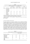
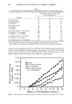
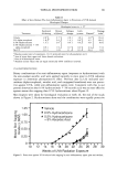
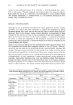
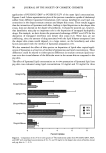
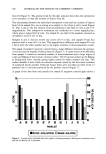
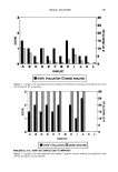
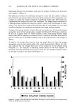
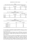
96 JOURNAL OF THE SOCIETY OF COSMETIC CHEMISTS carried out after addition of about 15 ml of Ecolite + (ICN Biomedical, Inc., Irvine, CA) to each system. The tape strippings and remaining skin were assayed as follows: Each sample was placed in a combustion cone and burned in a tissue oxidizer (Model 306, Packard Instrument Co., Downers Grove, IL). The separated radionuclides were assayed using a scintillation counter. RESULTS AND DISCUSSION Although the use of liposomal formulations for topical application has been steadily increasing, few studies have been undertaken to explain the mechanism by which liposomes deposit their lipids into the skin and the depth to which these lipids are deposited. Most in vitro transport studies concern themselves with permeation of drug through the skin and do not focus on the accumulation of lipid and entrapped ingre- dients in the various skin strata. In the cosmetic industry, it is essential to determine the extent to which the components of the formulation accumulate in the stratum corneum and, more importantly, to determine whether or not they are transported to the living epidermis and beyond. There is a vast literature dealing with the mechanisms by which liposomes interact with cell membranes and deposit their entrapped materials in the cell interior. However, little work has been done on the interactions between topically applied liposomes and the skin. The barrier function of the skin resides mainly in the stratum corneum, which lacks nuclei and organella but contains keratin fibers and a complex mixture of lipids. The stratum corneum of humans, mice, and pigs has been shown to be essentially devoid of phospholipids. Its lipid composition is rather non-polar and consists primarily of ceramides, triglycerides, cholesterol, fatty acids, and cholesteryl sulfate. These lipids are arranged in bilayer structures that fill the intercellular space in the stratum corneum. The primary pathway to the transport of water and other molecules is believed to reside mainly in these structures. The removal of these bilayer sheets either by solvent treat- Table I Distribution of Cholesterol (expressed as percent formulation applied + standard deviation) in Various Strata of Hairless Mouse and Pig Skin 24 hr After In Vitro Topical Application of Various PC/CH/CS Liposomal Formulations Onto Full-Thickness Skin (n = 4-5) REV DRV MLV Compartment Mouse Pig Mouse Pig Mouse Pig Total donor 29.6 --- 0.03 41.4 +- 8.03 64.3 + 5.6 42.0 + 7.6 49.0 --- 8.9 46.6 +- 4.2 Surface stratum corneum 48.2 -+ 6.8 25.3 +-- 4.9 18.6 -+ 3.9 26.4 -+ 2.8 27.1 + 3.3 22.9 -+ 3.8 Deeper stratum corneum 21.6 -+ 4.2 27.8 + 5.8 16.7 -+ 4.3 25.7 + 5.8 22.2 -+ 6.5 22.0 + 0.8 Deeper skin strata 1.2 -+ 0.3 5.5 + 0.7 1.4 + 1.3 5.8 + 0.5 1.6 -+ 0.2 8.3 + 1.6 Total skin 70.3 + 2.4 58.6 -+ 11.5 35.6 + 6.2 57.9 -+ 8.0 50.9 + 9.6 53.2 -+ 6.2 Receiver 0.1 -+ 0.05 0.05 -+ 0.05 1.1 + 0.02 0.1 -+ 0.1 1.2 + 0.03 0.2 + 0.06 All values were corrected to 100%.
Purchased for the exclusive use of nofirst nolast (unknown)
From: SCC Media Library & Resource Center (library.scconline.org)