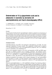
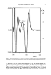
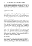
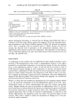
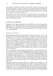
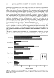
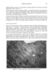
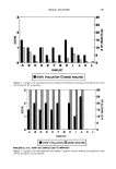
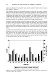
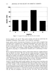
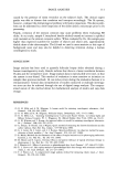
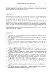
J. Soc. Cosmet. Chem., 43, 101-112 (March/April 1992) Quantitative assessment of cyanoacrylate follicular biopsies by image analysis DAVID G. GROH, OTTO H. MILLS, JR., and ALBERT M. KLIGMAN, Skin Care Laboratory, Personal Care Products Research and Development, Amway Corporation, Ada, MI 49355 (D.G.G.), Hill Top Research, Inc., East Brunswick, NJ 08816 (O.H.M.), and University of Pennsylvania, Philadelphia, PA 19104 (A. M. K. ). Received November 27, I99I. Presented at the Annual Scientific Meeting of the Society of Cosmetic Chemists, New York, December 5-6, 199•. Synopsis A method has been developed for quantifying the comedogenic effect of topically applied products using image analysis. Cyanoacrylate follicular biopsies are evaluated under polarized light, which brightly illu- minates horny casts and microcomedones, enabling rapid measurement of density and size distribution. A number of substances were tested on the backs of human subjects. There was a high correlation between stereomicroscope scores for comedogenicity and the readout by digital image analysis (r) 0.9). INTRODUCTION The rabbit ear model was the first widely used procedure for determining whether a topically applied substance could induce comedones. In the early eighties Mills and Kligman developed a human model for assessing comedogenic substances (1). The "non-animal" aspect of this model has greater meaning today than when it was first developed. In that model, test substances were applied under occlusion for one month on the upper part of the backs of young adult black men having large follicles. The suitability of each subject was checked by the noninvasive "follicular biopsy" technique (2). The follicular biopsy (FB) samples the contents of sebaceous follicles. To obtain a follicular biopsy, the skin surface was coated with a thin layer of methyl cyanoacrylate glue and a glass slide was applied firmly. After one minute, the slide was carefully peeled off, bringing with it a thin layer of stratum corneum and follicular horny extensions. Hyperkeratotic follicles on the follicular biopsy appeared as cylinders of horny material surrounding extracted veilus hairs (Figure 1). Follicular biopsies obtained at the end of the four weeks were then evaluated using a standard four-point scale: 0, noncomedogenic 1, smallish horny cylinders, involving at least half of the follicles 2, moderately sized horny masses lol

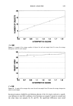
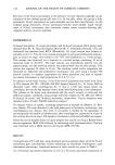
102 JOURNAL OF THE SOCIETY OF COSMETIC CHEMISTS •OLLICUL•R BIOPSY l•kV oe$D x155 D= 1 :•mm P = •.6T Figure 1. An ESEM (environmental scanning electron micrograph) of a follicular biopsy. over most of the field and 3, larger, globoid microcomedones over the entire field. This study showed that, although the human model was less sensitive, there was reasonably good correlation between the human and rabbit ear models. Recently, Mills et al. (3) compared black males and white females and found a good correlation between micro- comedone formation on the backs of these two groups. Image analysis (IA) has been used to evaluate skin surface topography (5-10), histology (11-14), and more recently, facial comedones by porphyrin fluorescence (15). We evaluated follicular biopsies by image analysis under polarized light. Polarized light has been effectively used for the photog- raphy of the skin (16) and consists of wavetrains whose planes of vibration are oriented in a parallel manner (17). METHODS AND MATERIALS CLINICAL STUDY The current study used a variation of the Mills and Kligman model. In the study the product was occluded on the upper backs of twelve white females with a history of acne. Each panelist was screened using the "follicular biopsy" technique, and a score of 1 or greater was required for inclusion in the test. The test was concluded in October of 1990. The test samples were: (A) a cleansing masque (B) a moisturizer (C) a positive control, acetylated lanolin alcohol (Acetulan©) and (D) a negative (no product) control. Test samples A and B were chosen because they were originally tested using the modified
Purchased for the exclusive use of nofirst nolast (unknown)
From: SCC Media Library & Resource Center (library.scconline.org)